Lymphatic malformations (synonyms and inclusions: cavernous lymphangioma; lymphangioma circumscriptum; macrocystic and microcystic lymphatic malformations)
LAST UPDATED: Mar 26, 2025
Introduction
Lymphatic malformations (LMs) are rare, sporadic, focal lesions composed of dilated lymphatic channels disconnected from the lymphatic system. Lesions can be macrocystic, microcystic, or combined.
This chapter is set out as follows:
History
- LMs are formed in embryo, but not all LMs are visible at birth
- While many are apparent by two years of age, others do not present until puberty, pregnancy or after trauma
Clinical findings
Distribution
- Lesions can occur at any site, although are most common on the neck, chest wall, and axilla
- While usually localised, LMs can occasionally be widespread (diffuse) - when lymphatic malformations are widespread in bone and soft tissue, the condition may be referred to as lymphangiomatosis
Morphology
- LM can be made up of macrocysts (large pockets containing lymph), or microcysts (very small pockets of lymph) - many LMs have both
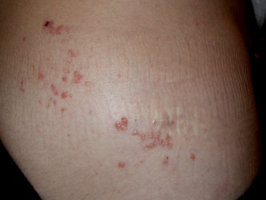
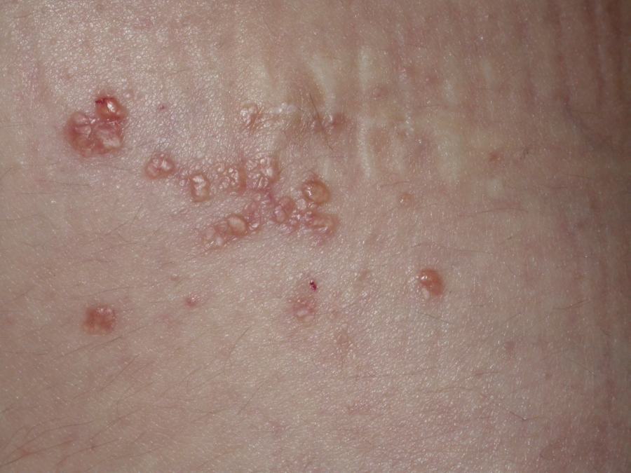
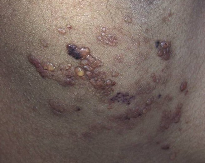
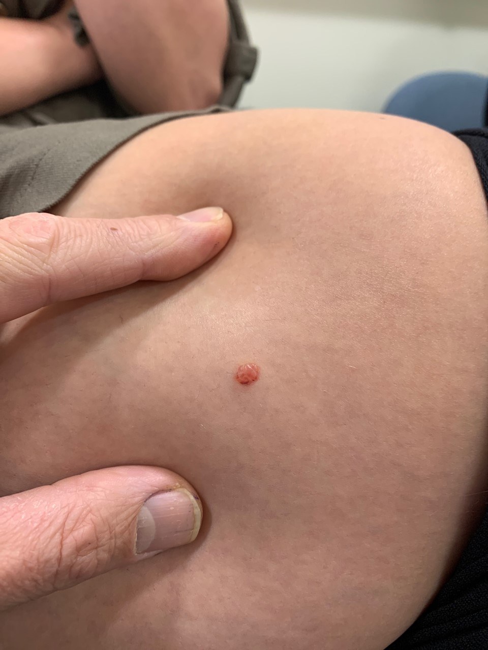
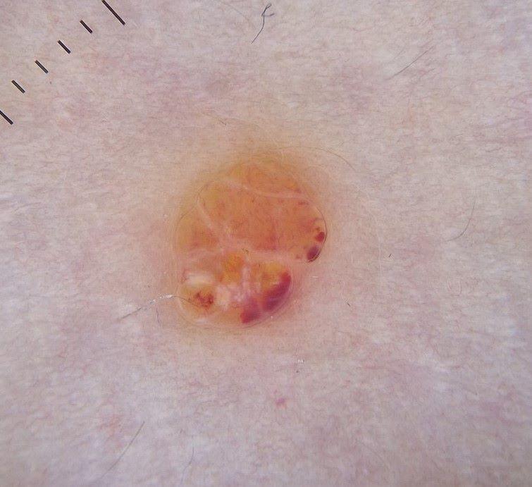
- Multilobulated, well‐defined lesions that are translucent, soft, but only slightly compressible on palpation, often with a frogspawn-like appearance
- Lesions can be clear, but become blue-purple when intracystic bleeding occurs
- Dermoscopically typical clods have the hypopyon sign with a two-tone appearance having a darker red or bluish colour at the base and a lighter yellow or pink colour at the top. The clods are separated by pale septae. Sometimes individual vessels can be seen feeding the clods
- Predominantly microcystic lesions are mostly located on the head and neck arising as ill‐defined vesicular plaques, which often invade adjacent structures
- Deeper involvement of the lymph system can occur that can be associated with lymphoedema
Associations
- Intralesional bleeding or infection can give rise to sudden pain - recurrent erysipelas/cellulitis are major complications that need to be treated rapidly as they can evolve into septicaemia
- Overgrowth (hypertrophy) and swelling of any affected area including the lips, tongue, jaws, cheeks, arms, legs, fingers, or toes
- Extensive LMs can be associated with venous malformations
- Visceral LMs can cause protein‐losing enteropathy and hypoalbuminaemia
- Various rare syndromes, eg, the Gorham–Stout syndrome, a bone disorder characterised by bone loss (osteolysis) associated with proliferation of lymphatic and/or other vessels
Clinical Images
Please refer to notes on image rights at bottom of the page with regards to individual image
ownership.
Investigations
- Doppler ultrasound (and sometimes MRI) is the best examination to confirm the cystic nature of this slow‐flow vascular malformation
Management
- Treatment is directed towards the specific symptoms that are apparent in each individual
- Uncomplicated cases lesions can often be observed
- Bacterial infection should be managed with systemic antibiotics and analgesia
- For problematic lesions options include:
- Macrocystic lesions can be treated by aspiration followed by sclerotherapy
- Cautery, laser, or radiofrequency ablation can be used to destroy superficial components, but recurrence may occur
- Excision, although recurrence is frequent with microcystic lesions
- The drugs Sildenafil and Sirolimus (rapamycin) have been found successful in some patients
Disclaimer - the author PCDS cannot accept responsibility for any misleading or incorrect statements, and the management of individual patients remains the direct responsibility of the individual doctor. We do however hope that visitors to this site can contact us regarding comments that are considered misleading or incorrect so that we can continue to improve the site.
Image Rights - The PCDS would like to thank Dermatoweb, DermQuest (Galderma), and others who have contributed images. All named individuals and organisations maintain copyright for the relevant images.