Mucinoses
LAST UPDATED: Jul 25, 2021
Introduction
Mucin (syn. protein–hyaluronic acid complex) is a normal component of the dermal extracellular matrix produced in small amounts by fibroblasts. The cutaneous mucinoses are a heterogeneous group of uncommon conditions whose main characteristic is abnormal mucin deposition in the skin, which may be localised or widespread. Mucin is capable of absorbing 1000 times its own weight in water, consequently in the mucinoses, where mucin is increased, the characteristic features may include oedematous and shiny skin.
This chapter, which is set out as below, provides a brief overview of the mucinoses:
Aetiology
The cause is largely unknown. The cutaneous mucinoses are divided into two categories:
- Primary cutaneous mucinoses in which the mucin deposit is the main histological feature resulting in recognised cutaneous lesions. Primary mucinoses can be divided into dermal and follicular mucinoses
- Dermal mucinoses - include lichen myxoedematosus, reticular erythematous mucinosis, scleredema, mucinoses in thyroid disease, papular and nodular mucinosis in connective tissue disease, self‐healing juvenile cutaneous mucinosis, cutaneous focal mucinosis, and digital myxoid cysts
- Follicular mucinoses - mucin accumulates in the epithelial hair follicle sheaths and sebaceous glands. The most common of these rare conditions is alopecia mucinosa (sometimes referred to as Pinkus follicular mucinosis). Urticaria‐like follicular mucinosis is extremely rare. Follicular mucinosis can also occur as a histological feature in cutaneous T‐cell lymphoma
- Secondary mucinoses refers to the histological findings of mucin resulting from other skin conditions. Clinically relevant lesions of mucinosis are absent. This is not discussed further
History
Localised lichen myxoedematosus (LM)
-
A rare condition with lesions limited to only a few sites. There are no sclerotic features, no paraproteinaemia, and no systemic involvement
-
Localised LM is classified into four subtypes:
-
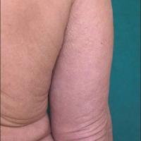
Acral persistent papular mucinosis: multiple ivory to skin‐coloured papules on the dorsal aspect of the hands and extensor surface of the distal forearms
-
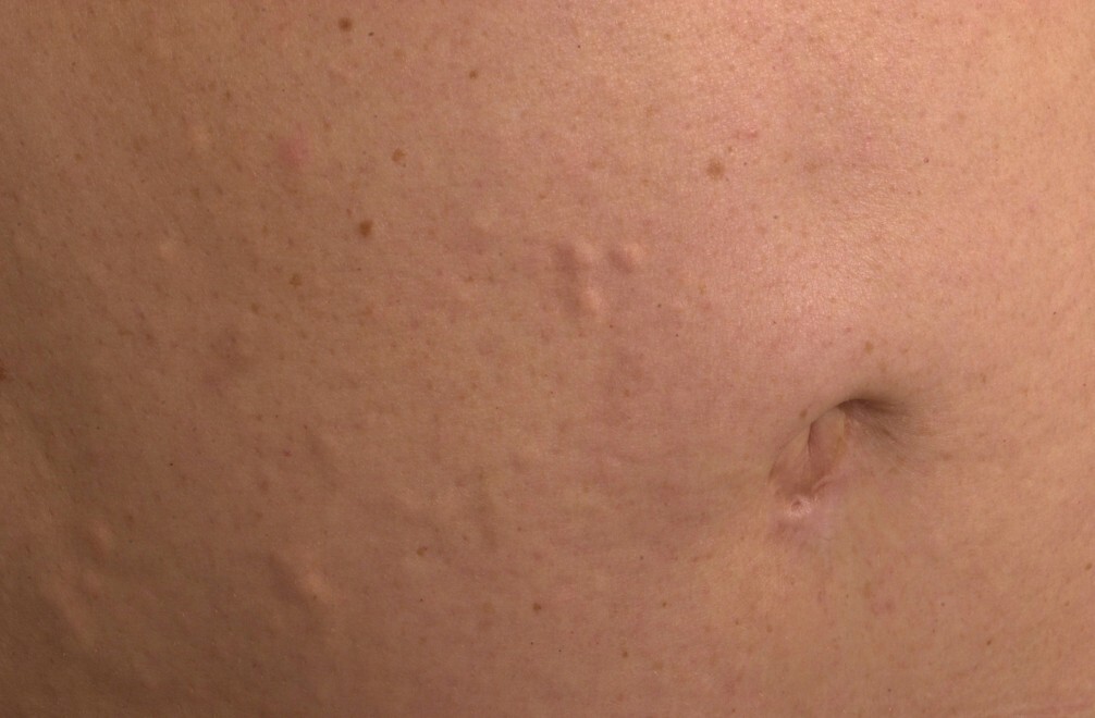
Discrete papular lichen myxoedematosus: a few to many, small, red-purple or skin‐coloured papules affecting the trunk and limbs in a symmetrical pattern
-
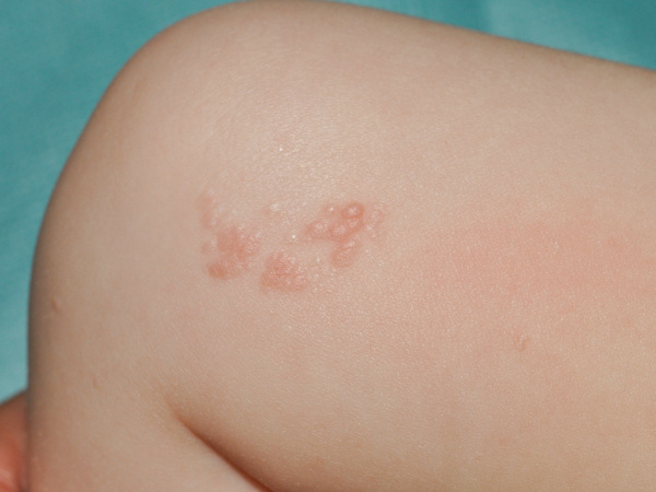
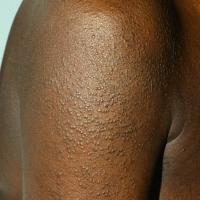
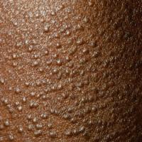
Cutaneous (papular) mucinosis of infancy: firm opalescent papules appear on the upper arms, neck and trunk
-
Nodular lichen myxoedematosus: multiple nodules on the limbs and trunk, with a mild or absent papular component
-
Spontaneous resolution of localised LM may occur
Scleromyxoedema
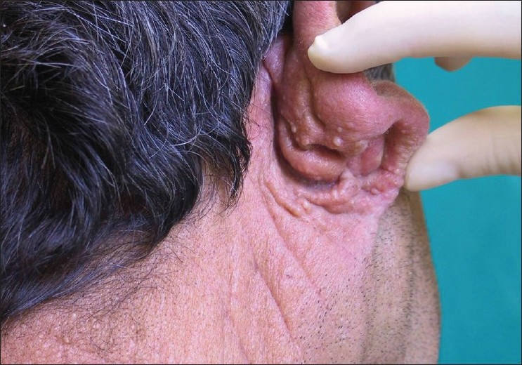
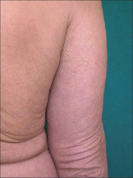
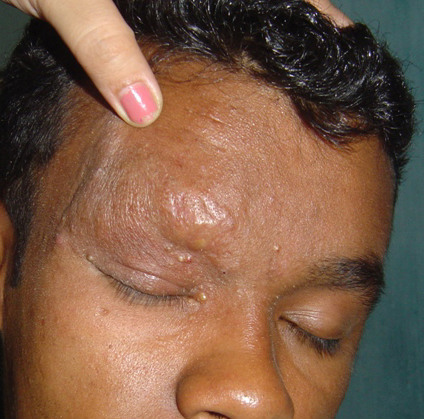
- A form of lichen myxoedematosus characterised by a generalised papular eruption on a sclerodermoid background
-
Is a rare disease that usually affects adults between the ages of 30 and 80 years
-
Distribution - a widespread eruption involving the upper limbs, head and neck, upper trunk and thighs
-
Morphology
-
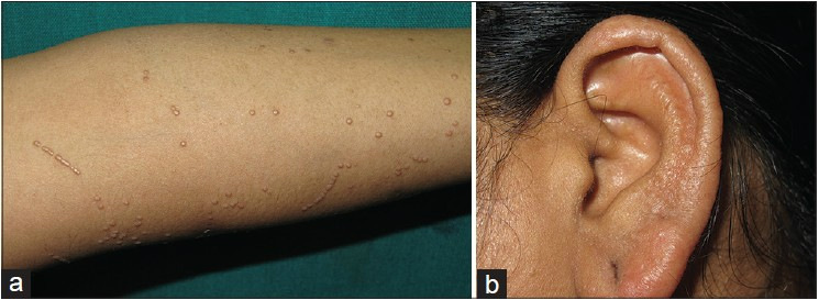
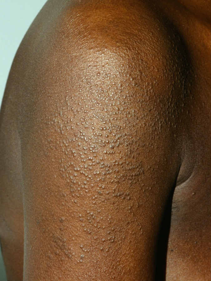
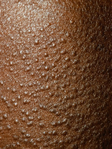
Many 2–3 mm, firm, waxy, closely spaced, dome-shaped or flat‐topped papules
-
Lesions are often arranged in a strikingly linear pattern
-
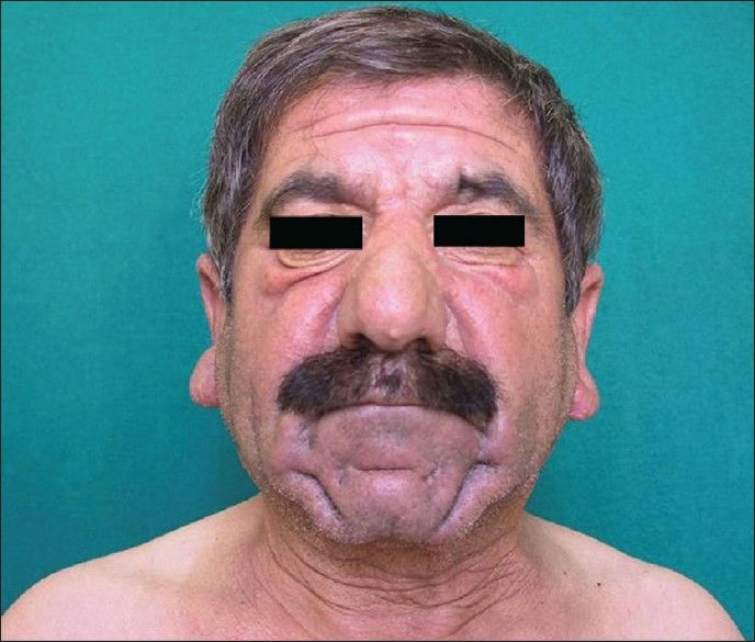
The skin is shiny and thick (ie sclerodermoid) and may be red-brown in colour
-
The glabella typically is involved with deep longitudinal folding. Deep furrowing can also be evident on the trunk, shoulders and limbs
-
Eyebrow, axillary and pubic hair may be sparse
-
As the condition progresses there is reduced mobility of the joints and around the mouth
-
Associations - scleromyxoedema is nearly always associated with haematological disease, especially monoclonal gammopathy, and also myeloma, lymphoma and leukaemia. If haematological disease is not apparent at presentation patients will require monitoring (twice yearly) of FBC, immunoglobulins & plasma electrophoresis looking for abnormalities and also trends, with a low threshold for haematological referral and bone marrow biopsy
-
Complications - most of the major organs can be involved with serious consequences
Papular and nodular mucinosis in connective tissue conditions
-
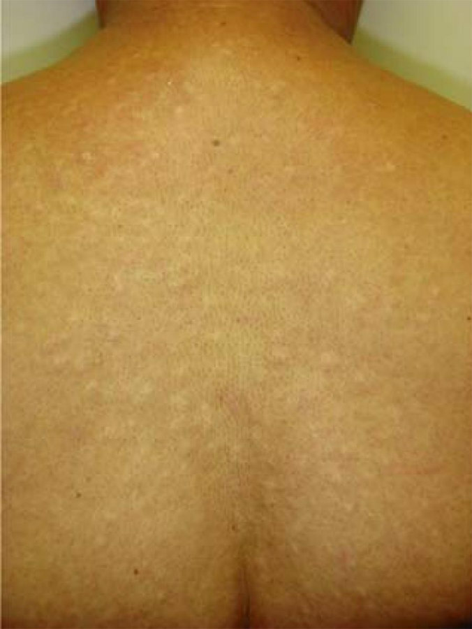
Skin‐coloured papules, nodules and plaque‐like lesions on the trunk and upper extremities may accompany or antedate a connective tissue condition, mostly lupus erythematosus, and rarely dermatomyositis or scleroderma
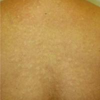
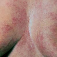
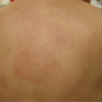
Reticular erythematous mucinosis
-
A rare condition predominantly affecting middle‐aged women, and generally not associated with systemic disease
-
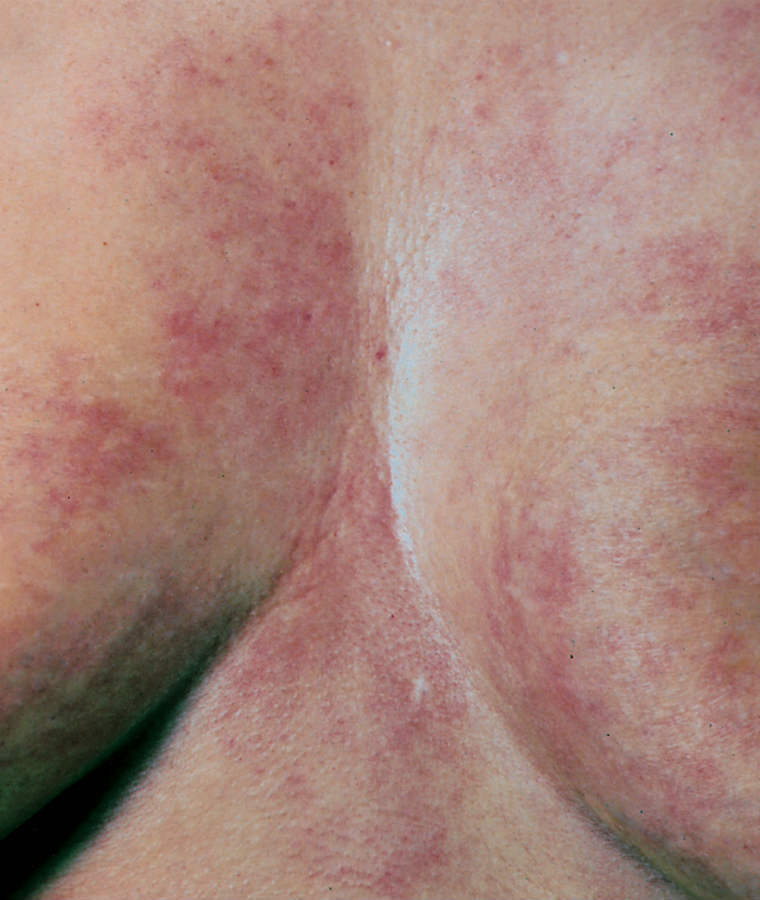
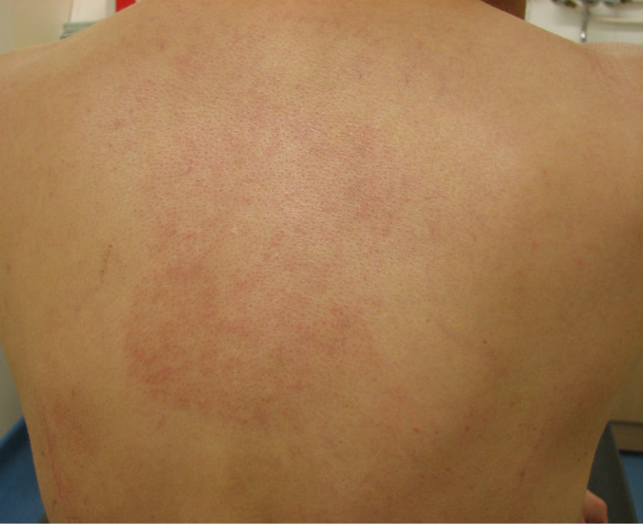
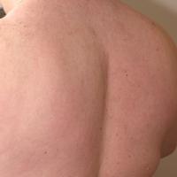
Distribution - midline of the chest or back, occasionally other sites
-
Morphology - erythematous macules and indurated papules or plaque‐like lesions with a reticular (net-like) pattern
Scleredema
Can present in two ways:
- Acute
- Mainly affects middle-aged women and children
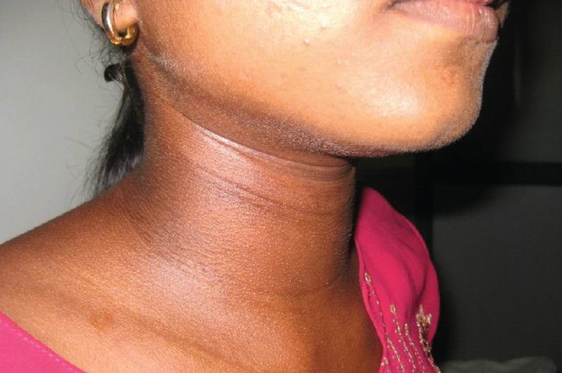
- Hardening of the skin of the face and neck develops quickly and spreads to the upper trunk and arms in a symmetrical fashion
- There can be loss of facial expression and difficulties in swallowing
- Hyperpigmentation is common feature
- This type of scleredema usually improves spontaneously over six months to two years
- Insidious onset
- A more insidious onset is associated with diabetes (sometimes referred to as scleredema adultorum of Buschke, or, scleredema diabeticorum), and sometimes a paraproteinaemia, which can be secondary to multiple myeloma. It can also be associated with other connective tissue conditions such as Sjögren's syndrome and rheumatoid arthritis
- Scleredema secondary to diabetes occurs particularly in adult men, and is very persistent
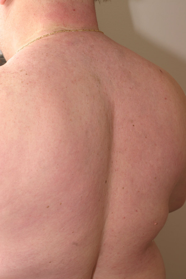
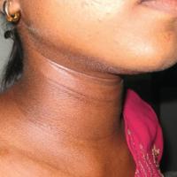
- The main sites affected are the skin of the neck and upper back that slowly thicken over months or years
-
For more information refer to the related chapter Scleredema
Pretibial myxoedema
-
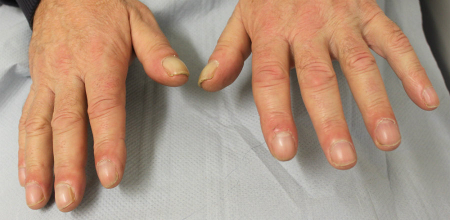
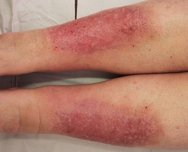
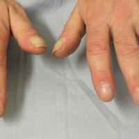
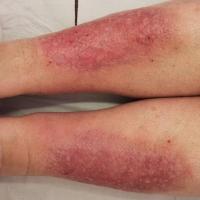
Pretibial myxoedema is one of the signs of Graves disease along with goitre, exophthalmos (prominent eyes due to deposition of myxoedema behind the orbit), thyroid acropachy (swelling of distal digits similar to clubbing, the nails may lift off the nail bed) and high circulating levels of long‐acting TSH
-
Less commonly, pretibial myxoedema has been described with Hashimoto thyroiditis and in patients with no history of thyroid dysfunction
-
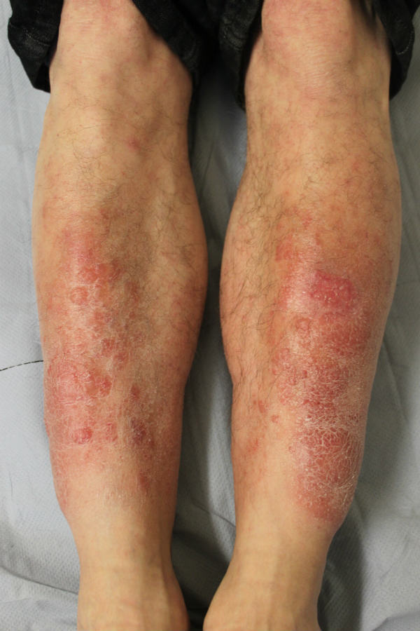
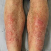
Distribution - shins and dorsa of the feet. Other sites are occasionally involved such as the thighs, upper extremities and face
-
Morphology
-
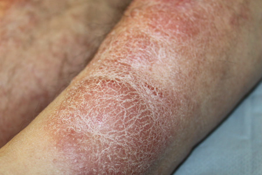
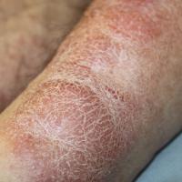
A diffuse skin thickening
-
There may be non‐pitting oedema, plaques or nodules
-
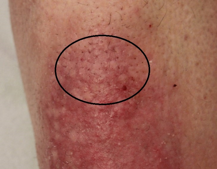
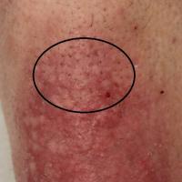
The skin can be yellow-brown, or exhibit a characteristic orange peel appearance and texture (peau d'orange) due to prominent hair follicles
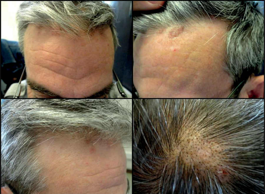
Alopecia mucinosa (benign follicular mucinosis) and folliculotropic mycosis fungoides (a cutaneous T‐cell lymphoma)
-
Alopecia mucinosa
-
Is the main type of follicular mucinosis
-
It is a rare condition that has a predilection for children, and adults in the third and fourth decades
-
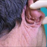
The most common sites involved are the scalp, face and neck, but any part of the body may be affected
-
Early signs of the condition are the presence of grouped follicular papules that appear in reddened plaques or patches. These are usually 2-5 cm in diameter but can be larger. One or more lesions may be present from onset or a single lesion may develop to multiple lesions over a few weeks or months. Hair loss is common from the affected follicles, in the early stages this is reversible ie the hair will grow back if the condition clears up, but in more severe cases complete follicular destruction prevents normal hair growth even if the inflammatory skin disease is then controlled
-
Folliculotropic mycosis fungoides
-
This is characterised by a more generalised chronic form in a slightly older age group, with larger and more numerous plaques on the extremities, trunk and face
-
It is probably best regarded as a follicular mucinosis associated with cutaneous T‐cell lymphoma rather than a primary condition
Other mucinoses
-
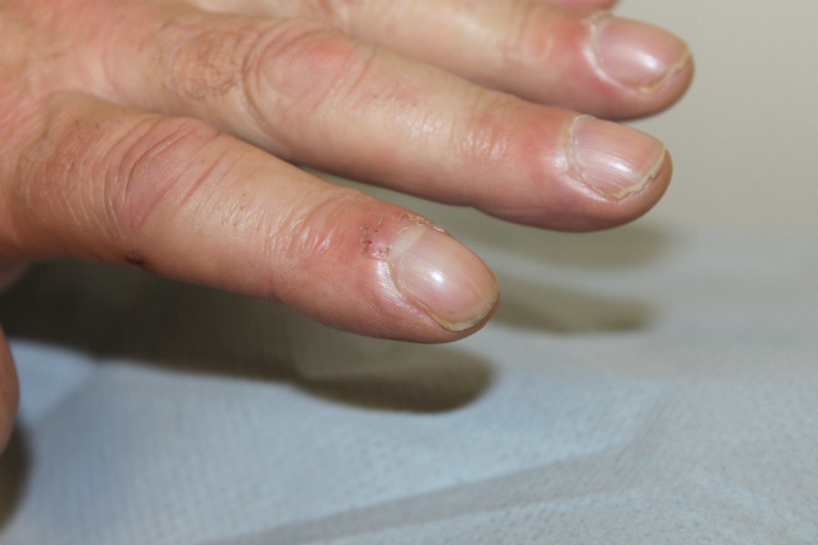
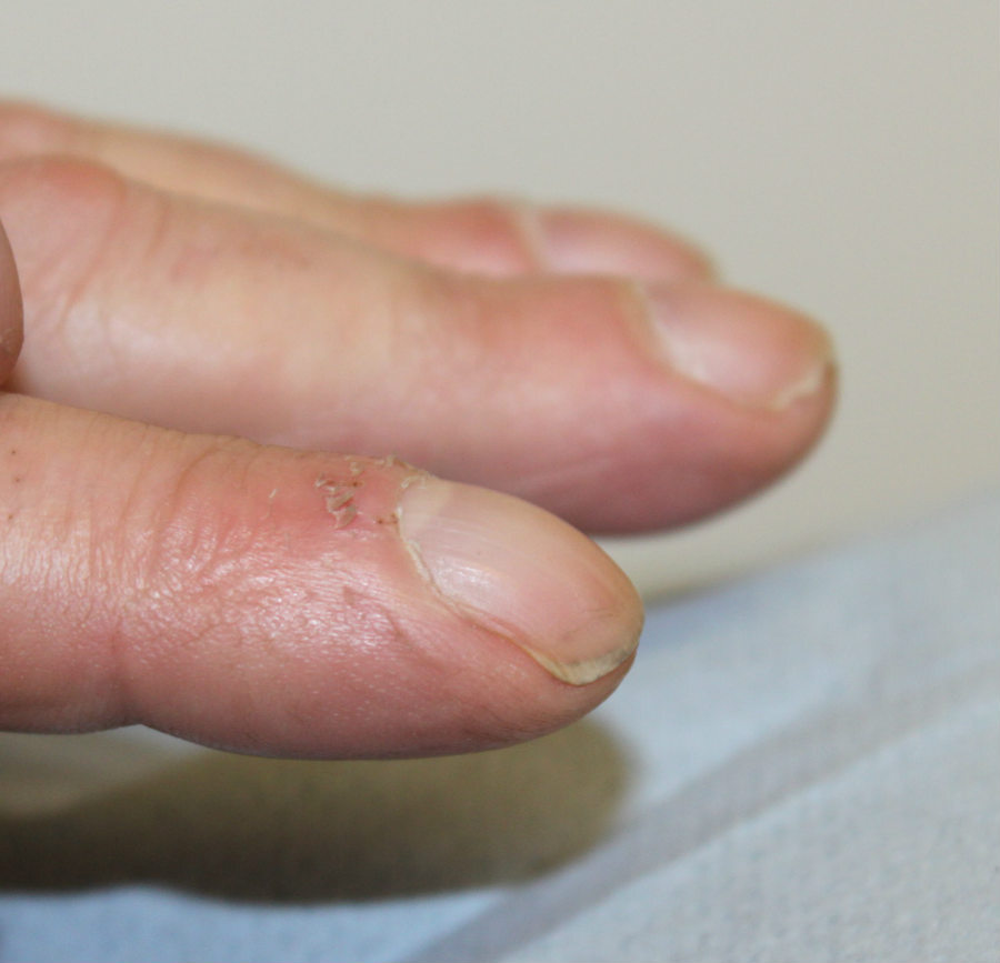
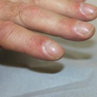
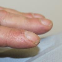
Digital myxoid cyst: a common disorder, found around the proximal nail fold. The cyst has a smooth shiny surface, and a clear jelly-like fluid may be expressed. As a result of pressure the cyst often causes a lateral groove in the nail, a few mm across, which extends the length of the nail
-
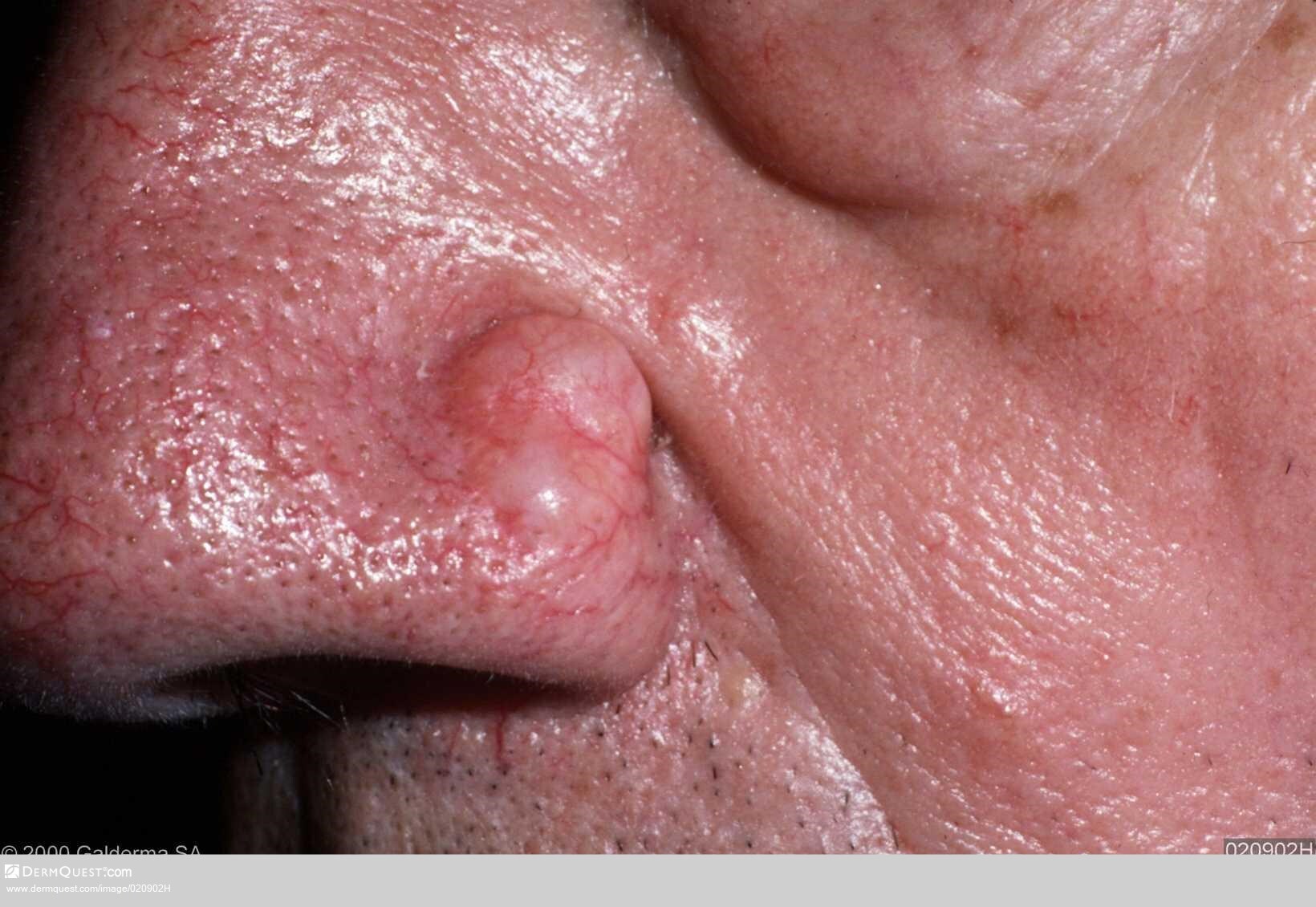
Cutaneous focal mucinosis: an asymptomatic, solitary, skin‐coloured papule or nodule, sometimes with a cystic appearance, that can occur anywhere on the body but not in proximity to the joints of the hands, wrists or feet
-
Self‐healing cutaneous mucinosis: affects young people with transient cutaneous lesions and mild inflammatory symptoms
Clinical Images
Please refer to notes on image rights at bottom of the page with regards to individual image
ownership.
1
2
3
4
5
6
7
8
9
10
11
12
13
14
15
16
17
18
19
20
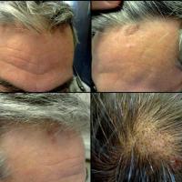
21
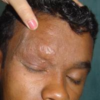
22
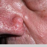
23
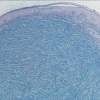
24
Investigations
-
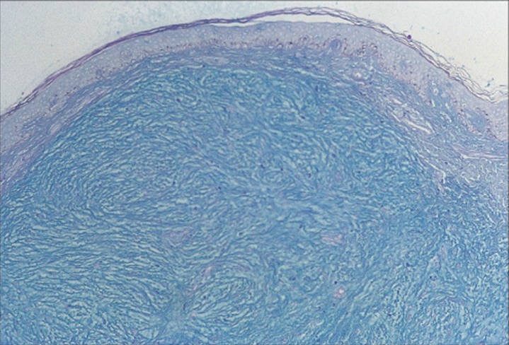
A skin biopsy is required - special stains such as the Alcian blue stain are used to detect the presence of increased mucin
Management
-
Patient with suspected mucinosis should be referred to a specialist
Disclaimer - the author PCDS cannot accept responsibility for any misleading or incorrect statements, and the management of individual patients remains the direct responsibility of the individual doctor. We do however hope that visitors to this site can contact us regarding comments that are considered misleading or incorrect so that we can continue to improve the site.
Image Rights - The PCDS would like to thank Dermatoweb, DermQuest (Galderma), and others who have contributed images. All named individuals and organisations maintain copyright for the relevant images.