Verrucous epidermal naevi
LAST UPDATED: Sep 19, 2022
Introduction
This chapter discusses both epidermolytic and non-epidermolytic verrucous epidermal naevi and is set out as follows:
History
-
Verrucous epidermal naevi (VEN) affect upto 0.5% of the population, of which non-epidermolytic lesions account for the majority
-
Males and females are equally affected
-
VEN may be congenital, although in over 50% of cases the onset is after birth
Clinical findings
-
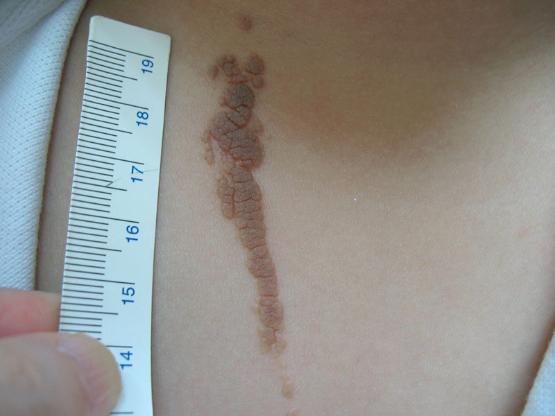
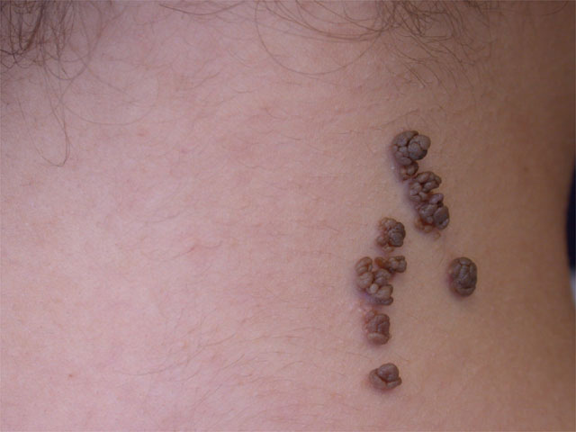
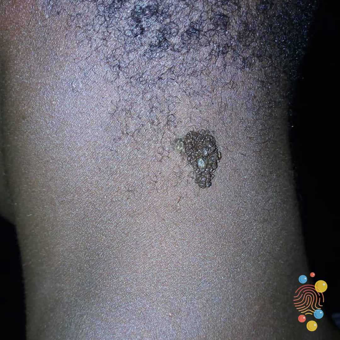
Non-epidermolytic and epidermolytic verrucous epidermal naevi have similar appearances
-
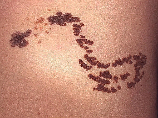
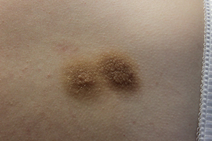
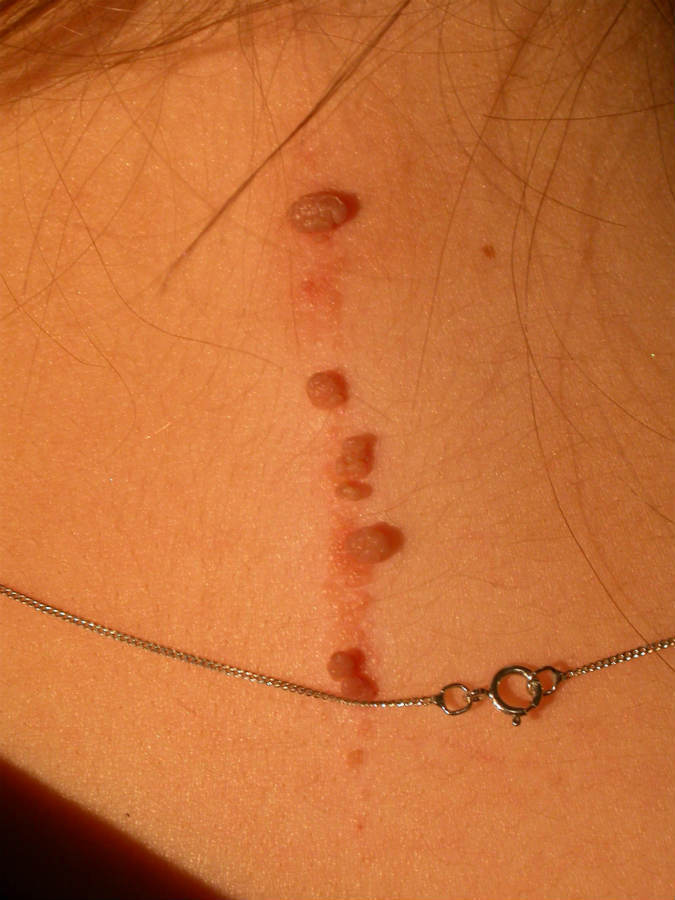
Arise as slightly pigmented, velvety or warty streaks / plaques
-
May be single or multiple
-
Follow Blaschko's lines, which are linear or spiral on the limbs and wave-like or v-shaped on the trunk - refer to the related chapter
-
Darken with age and may become more warty, sometimes with an erythematous base
-
Flexural lesions may be macerated
-
Extra-cutaneous features
-
Epidermal naevi represent mosaic mutations (in genes such as FGFR3, PIK3CA and HRAS) which can affect other tissues (including brain, eyes, bones) and can present as one of the ‘epidermal naevus syndromes'
Clinical Images
Please refer to notes on image rights at bottom of the page with regards to individual image
ownership.
Management
Step 1: are there any extra-cutaneous features?
- A small verrucous epidermal naevus (VEN) is very unlikely to be associated with extra-cutaneous abnormalities
- In larger lesions, any extra-cutaneous features are often (but not always) evident from a young age
Step 2: genetic tests
- Some patients with epidermolytic VEN run a risk of parenting children with a rare but serious condition known as epidermolytic ichthyosis (syn. bullous ichthyosiform erythroderma)
- One cannot differentiate clinically between epidermolytic and non-epidermolytic lesions, accordingly patients at a significantly increased risk of their offspring being born with epidermolytic ichthyosis require a biopsy (at an appropriate age) to look for histological confirmation of an epidermolytic VEN, and if positive need to be referred to genetics
- Those patients with a significantly increased risk include:
- if the epidermal naevus is large, near the gonads, or there are several blaschkoid segments involved
- if there is another relevant abnormality in the skin eg mosaic pigmentation
- if there are associated systemic features or learning disabilities
Step 3: treatment
- For many the result of treatment is disappointing
- Superficial treatments for smaller lesions, such as shave excision and cryotherapy, only bring temporary improvement
- Laser can be used on small and large lesions, but partial recurrence is common
- The size of many lesions makes surgical excision difficult
Disclaimer - the author PCDS cannot accept responsibility for any misleading or incorrect statements, and the management of individual patients remains the direct responsibility of the individual doctor. We do however hope that visitors to this site can contact us regarding comments that are considered misleading or incorrect so that we can continue to improve the site.
Image Rights - The PCDS would like to thank Dermatoweb, DermQuest (Galderma), and others who have contributed images. All named individuals and organisations maintain copyright for the relevant images.