Skin of colour - clinical variations
LAST UPDATED: Jul 01, 2021
Introduction
The term ‘pigmented skin’ refers to Fitzpatrick skin types IV to VI. This encompasses a wide range of ethnic and cultural groups, including Africans, African-Caribbeans, African-Americans, Hispanics, South East Asian, Chinese, Japanese and Native American Indians. Differences in cutaneous structure and function of pigmented skin, as well as cultural practices, can result in conditions with distinct presentations and varying prevalence that require unique treatment. This chapter is set out as follows:
Aetiology
-
All people have roughly the same number of melanocytes in skin tissue, regardless of colour. What differs is both the size and distribution of the melanosomes - increased levels of melanin and larger melanosomes provide greater photoprotection, resulting in a lower incidence of skin cancers. However, higher levels of melanin correlate with increased post-inflammatory hyperpigmentation ie darkening of the skin secondary to previous inflammation and trauma
Clinical findings
General problems related to skin of colour
-
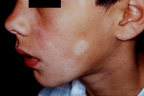
An increased incidence of eczema, pityriasis alba, tinea capitis, pseudofolliculitis, acne keloidalis nuchae, scars, acanthosis nigricans, lichen planus and fixed drug eruptions. Although sarcoid is an uncommon condition it is more prevalent in black skin and has many presentations
-
Post-inflammatory hyperpigmentation, sometimes post-inflammatory hypopigmentation, and the misuse of skin lightening agents are all more common
-
Although skin cancer is less common, the proportion of melanoma that is acral lentiginous melanoma in type is greater
-
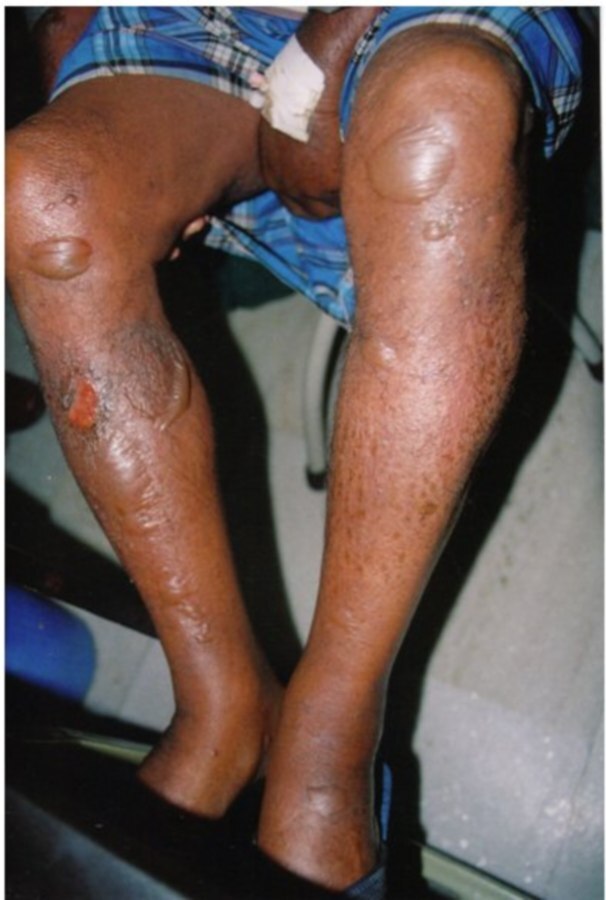
An increased awareness of tropical infection is required in immigrants arriving from certain parts of the world
-
Morphology
-
Increased pigmentation makes it difficult to recognise erythema
-
Surface scales takes on a grey appearance
-
Increased follicular involvement eg in eczema
-
More pronounced lichenification
-
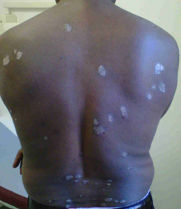
Psoriatic lesions can have a dark purple hue
-
Eczema can affect the extensor surfaces of the limbs, particularly at the elbows and knees
-
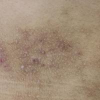
Scabies may have more in the way of pustules
Skin conditions that are more pronounced / problematic in skin of colour
Pigmentary demarcation lines
-
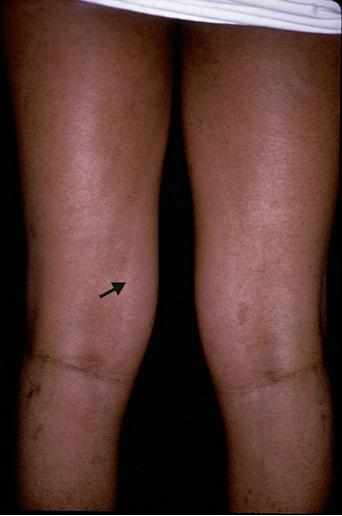
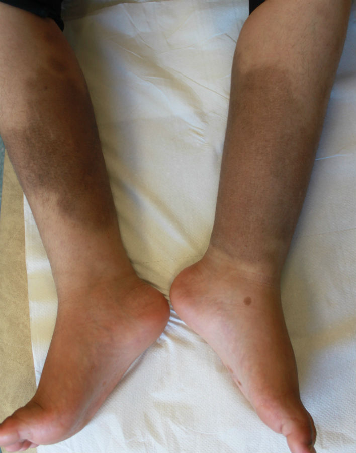
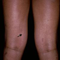
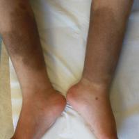
In all races, the dorsal skin of the limbs is darker than the ventral surfaces. This is more pronounced in pigmented skin, especially the anterolateral portion of the upper arm. These pigmentary demarcation lines (also known as Futcher’s or Voight’s lines) are seen in 20-30% of the black population
-
For more information refer to the chapter Pigmentary demarcation lines
Vitiligo
-
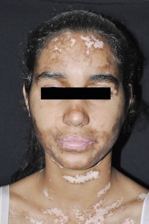
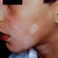
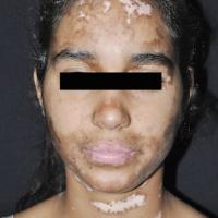
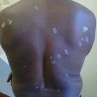
The contrast between normal skin and areas affected by vitiligo is more marked in persons with skin of colour, and as such the psychological impacts are often greater
-
If treatment is to be beneficial it needs to be initiated early - for more information refer to the chapter Vitiligo
Acne vulgaris
-
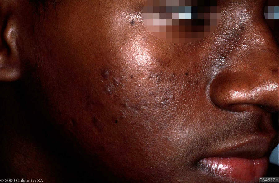
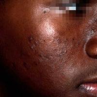
Acne is no more common or severe in skin of colour, but post-inflammatory hyperpigmentation can be significant and often persists for months to years. As such, early and more aggressive treatment is advocated, including early referral for consideration of isotretinoin
-
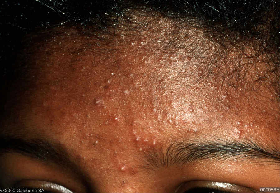
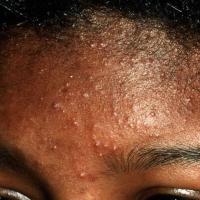
Secondary acne can be a result of cultural practices - hair oil can induce pomade acne on the forehead and skin lightening products containing topical steroids can cause a steroid-induced acne
-
Topical retinoids, benzoyl peroxide and azelaic acid play an important role in the treatment of the comedonal aspects of acne. If used daily they often cause dryness and irritation, which in skin of colour can lead to further hyperpigmentation. Accordingly these treatments should initially be used for shorter periods of time (eg in an evening and washed off before bed) and perhaps less frequently (eg every second or third day), if tolerated the duration and/or frequency of treatment can be increased
-
For more information refer to the chapter Acne vulgaris
Lichen planus pigmentosus
-
This variant of lichen planus may or may not be associated with typical LP papules and itch is often absent
-
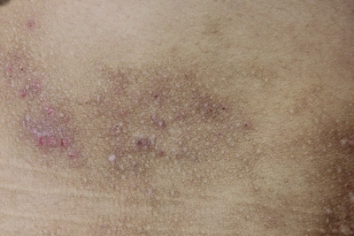
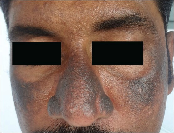
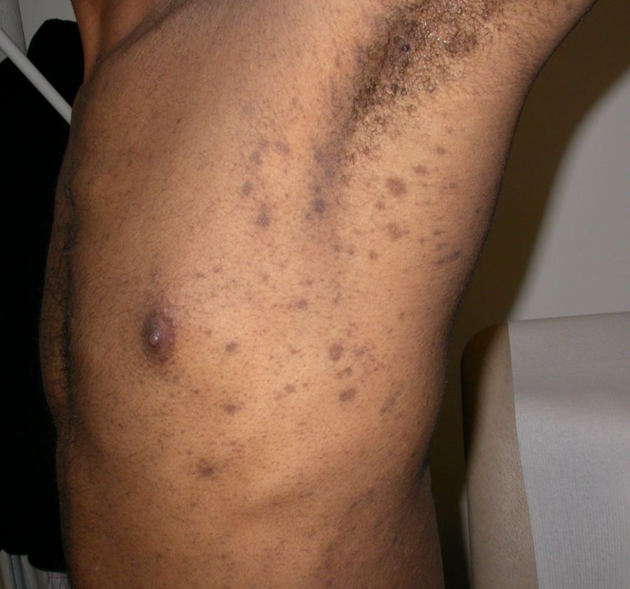
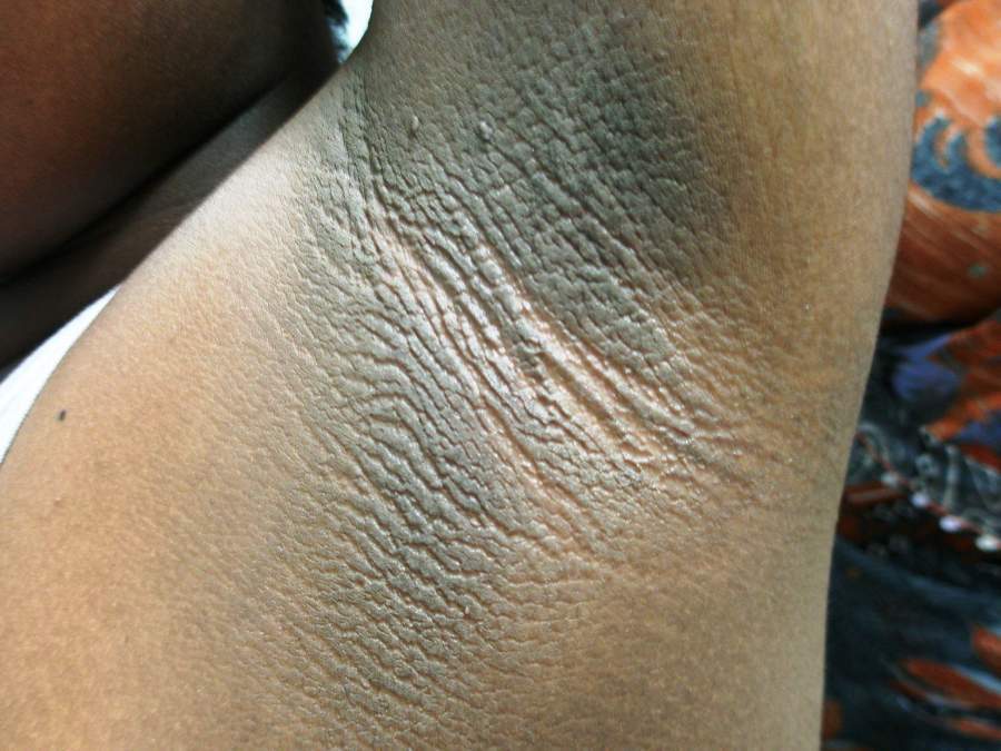
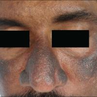
The macular hyperpigmentation involves chiefly the face, neck and upper limbs, although it can be more widespread, and varies from slate grey to brownish black. It is mostly diffuse, but reticular, blotchy and perifollicular forms are seen. Occasionally there is a striking predominance of lesions at intertriginous sites, especially the axillae
-
The duration ranges from two months to two years and there is no known effective treatment
-
For more information refer to the chapter Lichen planus
Post-inflammatory hyper- or hypopigmentation
-
Post-inflammatory hyperpigmentation (and less so hypopigmentation) is more common
-
It is usually caused by an inflammatory dermatosis and sometimes trauma
-
The situation can be made worse if the patient attempts to self-treat and apply various chemical bleaching substances (eg hydroquinone) to the skin, this is referred to as exogenous ochronosis
-
The main aim of treatment is to treat any on-going inflammation
-
Epidermal post-inflammatory hyperpigmentation will take 6-12 months to fade. Deeper pigment may take years. An important part of therapy is sunscreen with a minimum sun protection factor (SPF) of 30 to prevent darkening of the post-inflammatory hyperpigmentation. For post-inflammatory skin conditions of the face please refer above to the notes on acne vulgaris
- Phototherapy has been successfully used in some patients
-
Patients can also be referred for camouflage
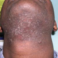
Pseudofolliculitis barbae
-
This is an inflammatory follicular condition with an estimated prevalence of 45-85% in African-Americans. Hispanic men are the next most-affected group
-
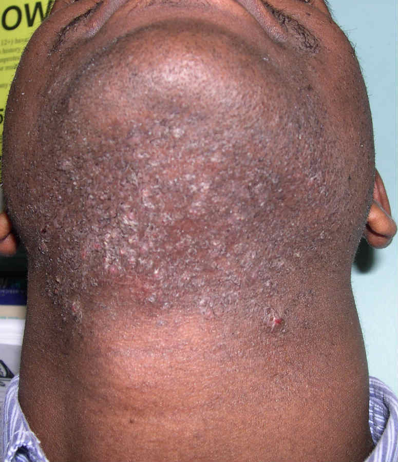
It is most common in men, affecting the beard area or neck, but can also affect women on areas of the body where hair removal practices have been used
-
It is thought that the natural tendency for curly hair to recoil in this ethnic group predisposes cut hairs to re-enter the skin. As the hair re-enters the dermis, it causes an inflammatory foreign body response, which manifests as papules and pustules
-
Hyperpigmentation and keloid scarring are frequent complications
-
For more information refer to the chapter Pseudofolliculitis barbae
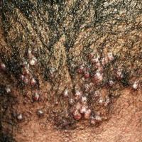
Acne keloidalis nuchae
-
Occurs in persons of all races but most commonly in men of African descent, followed by Hispanic, Asian, and white men. Persons affected are primarily post-pubertal, and onset is typically before 50 years of age
-
Close shaving and close-cropped hair are the main risk factors, but friction from shirt collars and helmets has also been implicated
-
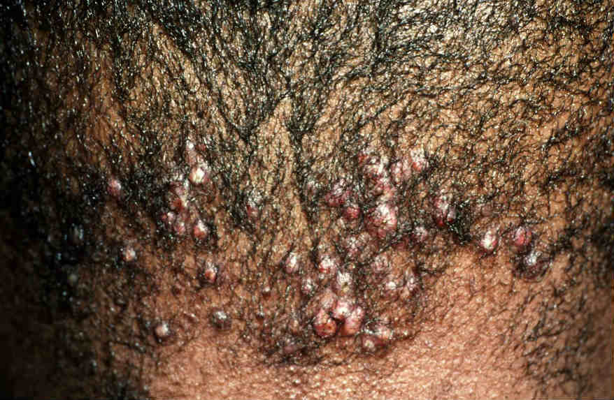
The condition begins as a chronic folliculitis of the posterior neck and occipital scalp, which progresses to keloidal papules and plaques causing a scarring alopecia - early treatment of Acne keloidalis nuchae correlates with a good prognosis
Scarring - keloid, hypertrophic and atrophic
-
There is a higher incidence in black skin, Hispanic persons, and Asians
-
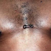
Keloids are benign growths that represent an overgrowth of scar tissue at sites of trauma, or sometimes arise spontaneously. Keloids are often confused with hypertrophic scars. Hypertrophic scars typically develop soon after the inciting trauma and are found on areas of the body with frequent motion, such as extensor surfaces. They are confined to the borders of initial injury and may regress. Conversely, keloids develop months to years after injury and most commonly arise on the ears, jaw line, nape of the neck, scalp, chest, and back. Keloids extend beyond the borders of original injury, often progressing over time
-
Refer to the chapter Keloid scars for more information
Alopecia
-
Hair care practices - although no biochemical differences of hair have been found among persons of different ethnicities, there are notable differences in the shape of the hair follicle, leading to differences in texture, fragility, and manageability of hair. Persons of African descent have the greatest degree of curl and increased fragility. Educating patients about avoiding traumatic hair practices is key in prevention
-
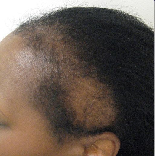
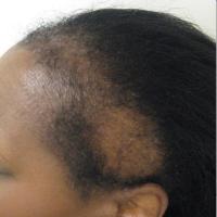
Traction alopecia - is the loss of hair from excessive pulling, and may result from tension placed on the hair with braids and weaves. Hair loss often occurs at the frontal and temporal hair lines. For more information refer to the chapter Traction alopecia
-
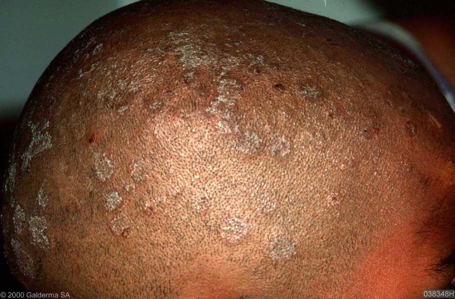
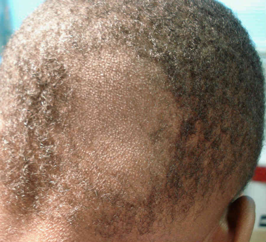
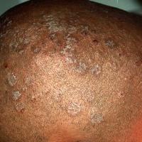
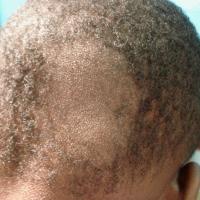
Tinea capitis - is an infection of hair follicles and surrounding skin with dermatophyte fungi and presents with inflammation, scaling and sometimes patchy hair loss. The condition is most commonly seen in African-Caribbean prepubertal children in urban communities. This is thought to be a result of hairstyling practices, such as close-cropped styles and tight plaits, which make the follicles more vulnerable to infection - patients are advised against sharing hair clippers because this can spread the infection. It is clear that the pattern of tinea capitis in the UK has changed in the past ten years with a significant rise in the incidence and prevalence of cases of infection due to Trichophyton tonsurans. For more information refer to the chapter Tinea capitis
-
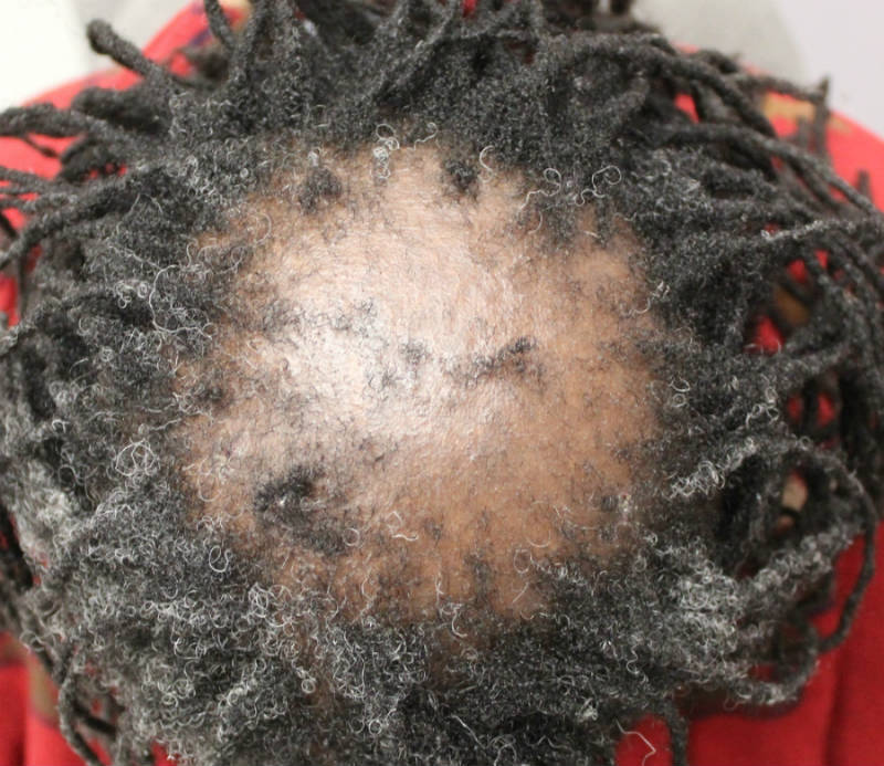
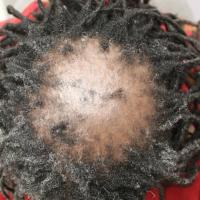
Central centrifugal cicatricial alopecia - a condition exclusively seen in Fitzpatrick type VI skin. It presents as a diffuse scarring alopecia that begins at the crown and spreads outwards, classically starting in the mid-30s. Women are more commonly affected than men. The aetiology is unknown, the initial hypotheses was that the condition was associated with heated styling instruments or chemical straighteners. Patients are encouraged to discontinue use of relaxers, limit traction, and decrease the amount of heat applied to the hair. No ideal treatment currently exists; however, options include topical and intralesional steroids with oral antibiotics. Early intervention is most effective. For more information refer to the chapter Central centrifugal cicatricial alopecia
Dermatosis papulosa nigra
-
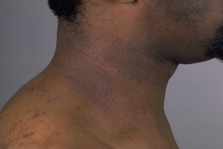
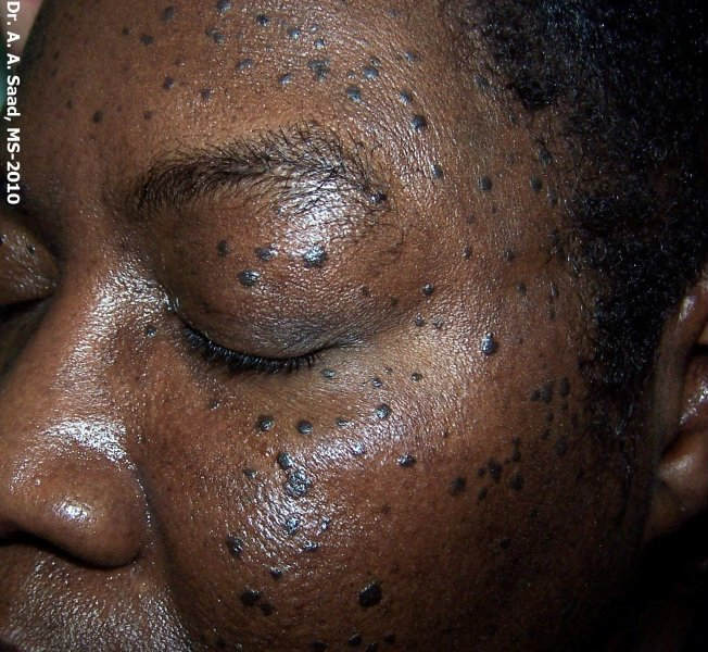
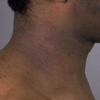
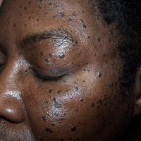
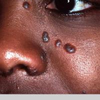
Is a common benign skin condition that occurs predominantly in skin of colour, presenting with multiple 1-5 mm multiple brown to dark-brown, dome-shaped papules predominantly on the face and neck
-
The highest prevalence is in black skin, reportedly between 10% and 30%. However, the condition also affects Asians, Hispanic persons, and whites. Onset is typically after puberty
-
Treatment is not usually required, and should be avoided if possible as the risks of any physical treatments include post-inflammatory hyper- or hypopigmentation
-
For more information refer to the chapter Dermatosis papulosa nigra
Skin cancer
-
Although skin cancer is relatively uncommon in skin of colour, patients are likely to present with more advanced skin cancer and have lower survival rates when compared to Caucasians
-
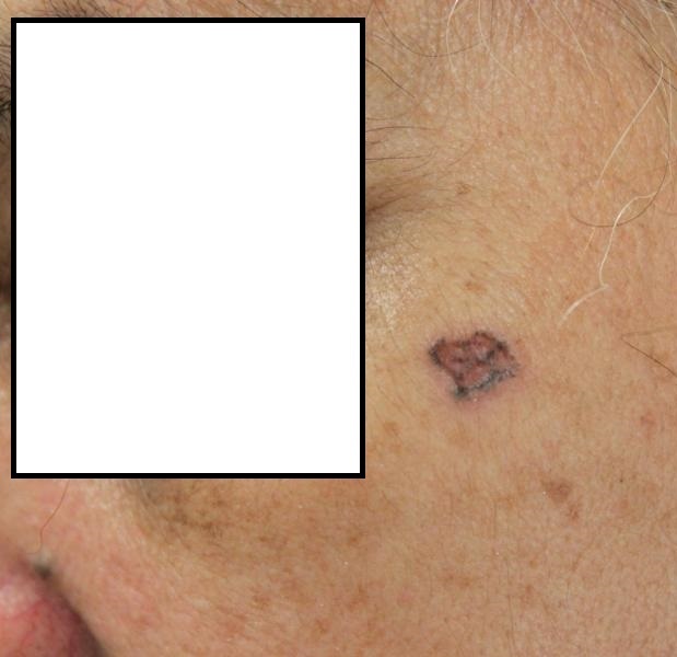
Basal cell carcinoma is often more pigmented than compared to lesions in Caucasian skin
-
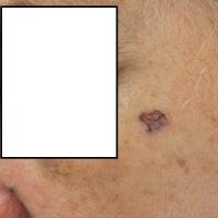
Squamous cell carcinoma is the most common skin cancer in black skin and Indians, and the second most common in Chinese, Japanese, and Hispanic persons. UV exposure is a known risk factor in Caucasians, with most lesions in sun-exposed areas. In comparison, the influence of UV light on the aetiology of squamous cell carcinoma in pigmented skin is unclear, and lesions commonly occur in non sun-exposed sites. The most important risk factors in pigmented skin is chronic inflammation and scarring
-
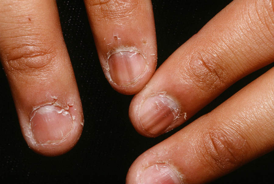
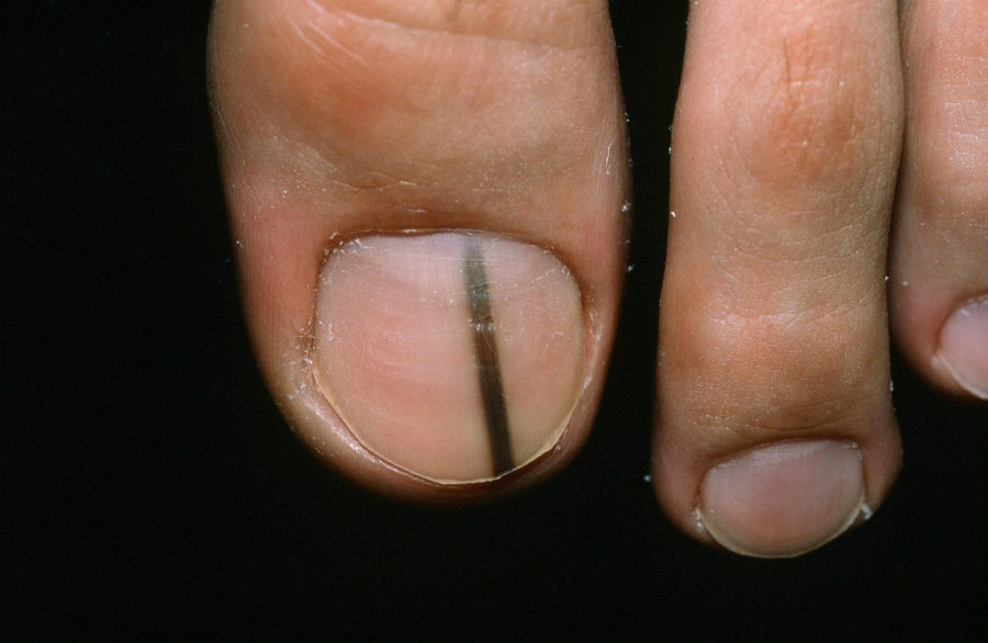
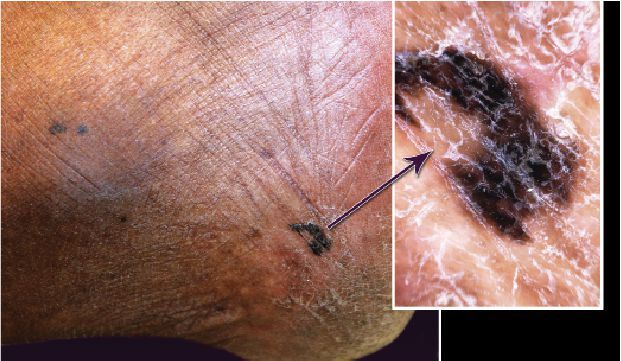
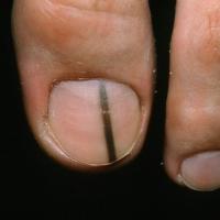
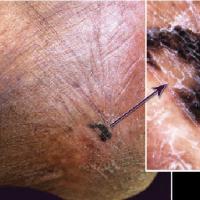
Acral lentiginous melanoma (melanoma involving the palms, soles, or nail beds) is the most common subtype of melanoma in black skin and Asians. Superficial spreading melanoma is the most common subtype in Hispanic persons and in whites. Evaluation of melanoma of the nail bed can be difficult because many persons with skin of colour have melanonychia (benign linear pigmentation of the nails) - this can be seen in childhood, but becomes more obvious with age. The width of pigmented bands varies from a few millimetres to the whole width of the nail and the colour varies from light brown to black. Pigmentation often affects multiple nails, which helps to distinguish it from subungual haemorrhage, melanocytic naevi and most importantly, melanoma. However, in adults any new or changing line in a nail (starting from the nail base) should be considered for an urgent referral (two-week wait) - for more information refer to the chapter Nail disorders
Tropical infection
Clinical Images
Please refer to notes on image rights at bottom of the page with regards to individual image
ownership.
1
2
3
4
5
6
7
8
9
10
11
12
13
14
15
16
17
18
19
20
21
22
23
24
25
26
27
Other resources
Disclaimer - the author PCDS cannot accept responsibility for any misleading or incorrect statements, and the management of individual patients remains the direct responsibility of the individual doctor. We do however hope that visitors to this site can contact us regarding comments that are considered misleading or incorrect so that we can continue to improve the site.
Image Rights - The PCDS would like to thank Dermatoweb, DermQuest (Galderma), and others who have contributed images. All named individuals and organisations maintain copyright for the relevant images.