Combined naevus

LAST UPDATED: Oct 03, 2025
Introduction

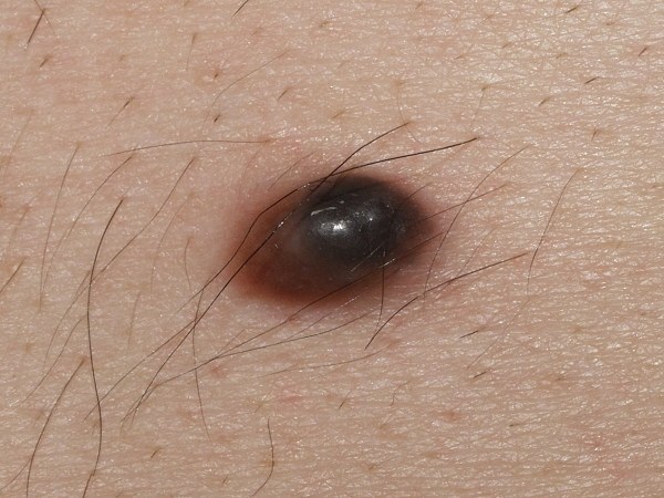
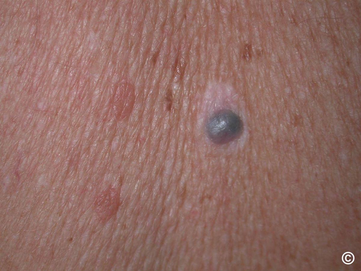
Combined naevi are clinically defined as melanocytic lesions comprising two or more distinct melanocytic naevus components; histologically lesions are determined by the presence of two or more different naevus cell types in one biopsy specimen. They are uncommon representing less than 1% of all biopsied melanocytic naevi. Combined naevi include any combination of melanocytic naevi, the most common is a blue naevus combined with a common brown melanocytic naevus. Given the combination of colours some combined naevi mimic melanoma.

This chapter is set out as follows:


History

  • Although combined naevi are mainly congenital, many are not visible at birth
  • Combined naevi can be seen at any age, most commonly presenting in young adults
  • There is a slight male predominance 

Clinical findings

  • Distribution
    • Most frequently affect the trunk and head and neck region,  less commonly the extremities 
       
  • Morphology
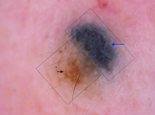
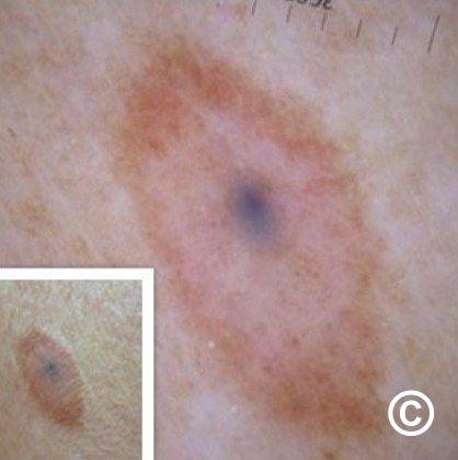
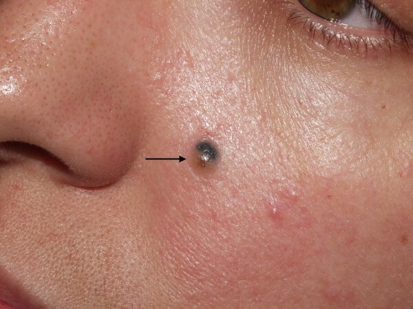
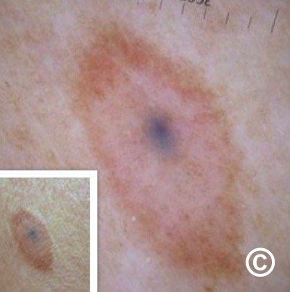
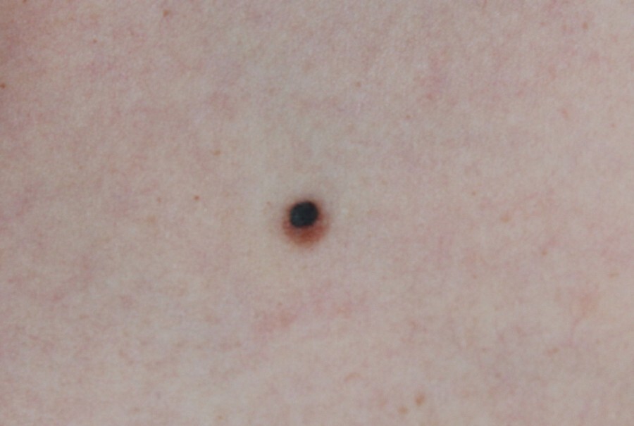
    • Since combined naevi are composed of distinct naevi, their clinical appearance could be very diverse. In practice, combined naevi are usually small, flat, or minimally raised lesions, often characterised by a small blue area (rarely black or white) in the context of a larger area of brown colour surrounding the blue naevus 
       
  • Dermoscopy (hover over terminologies for description)
     
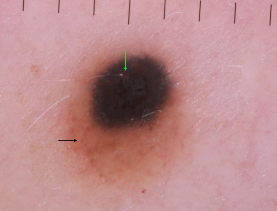
    • Since combined naevi are composed of at least two different naevus types in various combinations, their dermoscopic features are characterised by mostly multicomponent, unspecific, and sometimes random patterns
       
    • Differentiating combined naevi from melanoma:
       
      • Combined naevi - typically, the multicomponent structure of combined naevi exhibit a reasonably symmetrical appearance, which is consistent with already established findings that benign naevi tend to exhibit symmetry. The structureless blue part of the lesion is commonly central or off-centre, usually covering about 30% of the lesion. If located centrally, the blue structureless area is surrounded by another pattern, most commonly a brown globular pattern or pigment network. Benign lesions should lack dermoscopic melanoma-specific features
         
      • Melanoma - should be considered if the blue area is greater than 30% or touches the edge, if blue is combined with a colour other than brown, in chaotic lesions (asymmetry of structures or colour) and those with any dermoscopic melanoma-specific features

Clinical Images

Please refer to notes on image rights at bottom of the page with regards to individual image ownership.
Management

  • In cases of diagnostic uncertainty lesions should be referred on an urgent skin cancer pathway  
  • Lesions are often excised with a 2 mm margin of normal skin to confirm the diagnosis and exclude melanoma 

Disclaimer - the author PCDS cannot accept responsibility for any misleading or incorrect statements, and the management of individual patients remains the direct responsibility of the individual doctor. We do however hope that visitors to this site can contact us regarding comments that are considered misleading or incorrect so that we can continue to improve the site.

Image Rights - The PCDS would like to thank Dermatoweb, DermQuest (Galderma), and others who have contributed images. All named individuals and organisations maintain copyright for the relevant images.

Quick Links

The following pharmaceutical companies have had no involvement in the content of this website or in our conference programmes


Almirall
Galderma
Glenmark
Johnson & Johnson
La Roche-Posay
LEO Pharma
Pierre Fabre
Schuco