Blue naevus

LAST UPDATED: Oct 03, 2025
Introduction

A blue naevus is a benign (harmless) type of melanocytic naevus derived from spindle-shaped or, less commonly, ovoid naevus cells, located deep within the dermis. 

This chapter is set out as follows: 


Aetiology

Blue naevi are thought to arise during foetal life when melanocytes become arrested in the deep dermis during migration from the neural crest. The melanocytes of blue naevi are located deeper in the skin as compared to those of brown naevi


History

  • Twice as common in women; more prevalent among Asians
  • Can arise at any age; most commonly presenting in older children and young adults
  • Once evolved, blue naevi remain unchanged
  • Usually solitary
  • Multiple 'eruptive' blue naevi present as a sudden, rapid appearance of blue naevi at various anatomical sites, this rare phenomenon may be idiopathic, arise in pregnancy, or be secondary to sunburn, trauma, or other skin injury

Clinical findings

Distribution

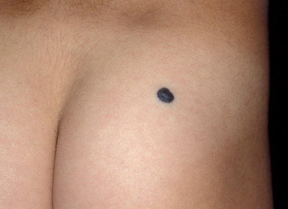
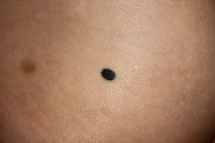
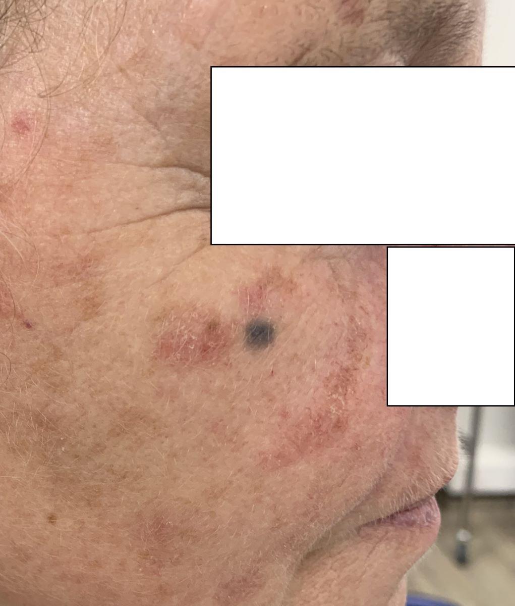
  • Can arise anywhere; most commonly the distal extremities (dorsum of hands and feet), buttocks, scalp and face 

Morphology 

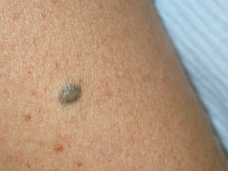
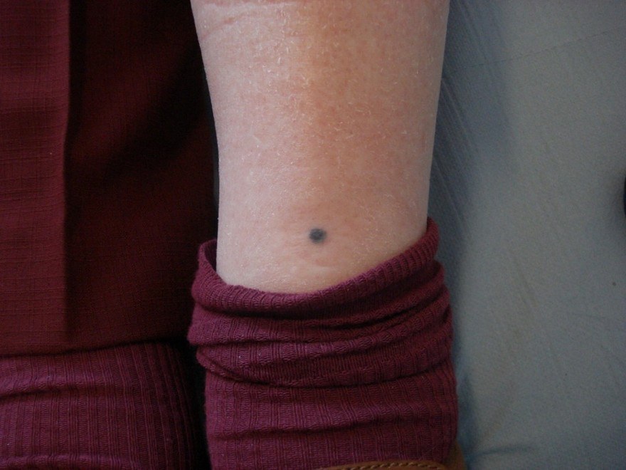
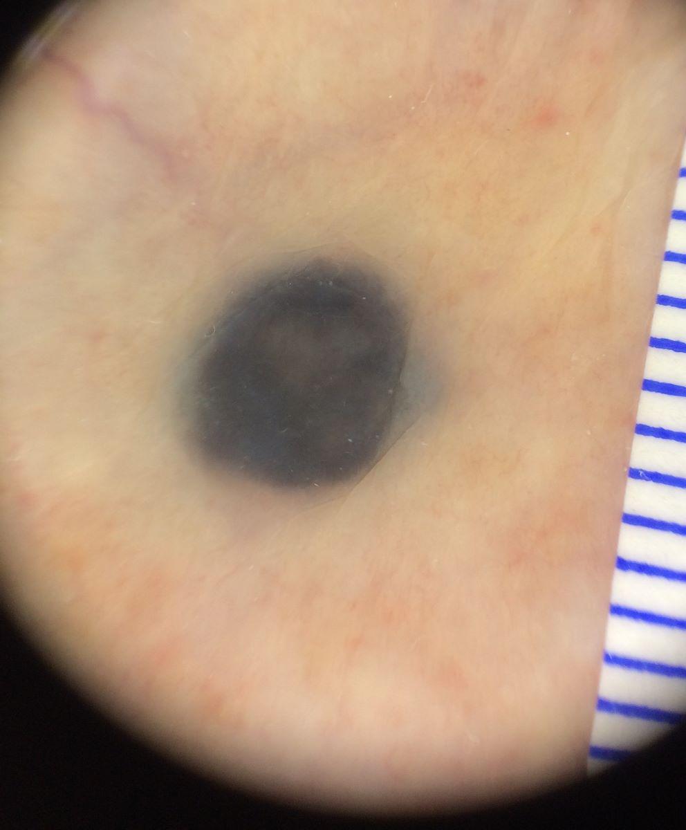
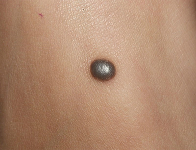
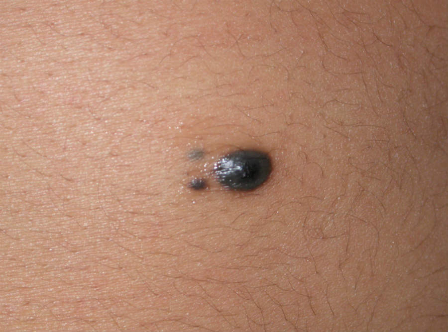
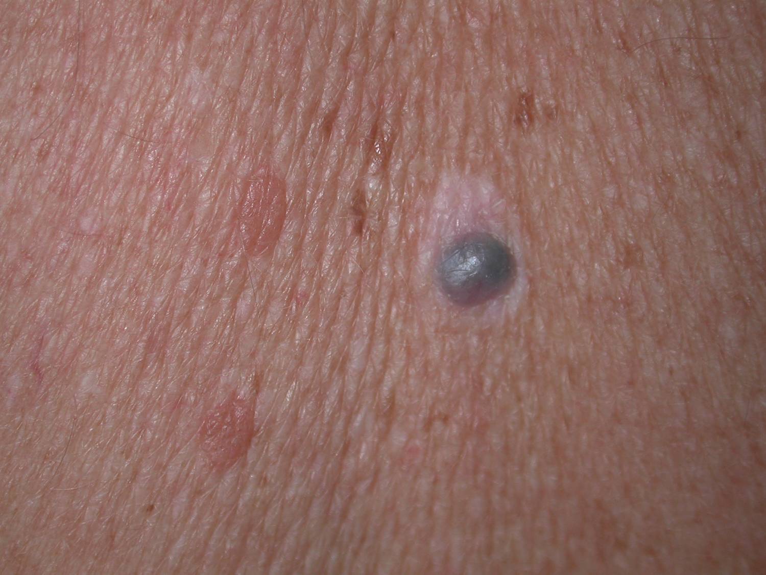
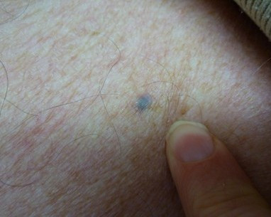
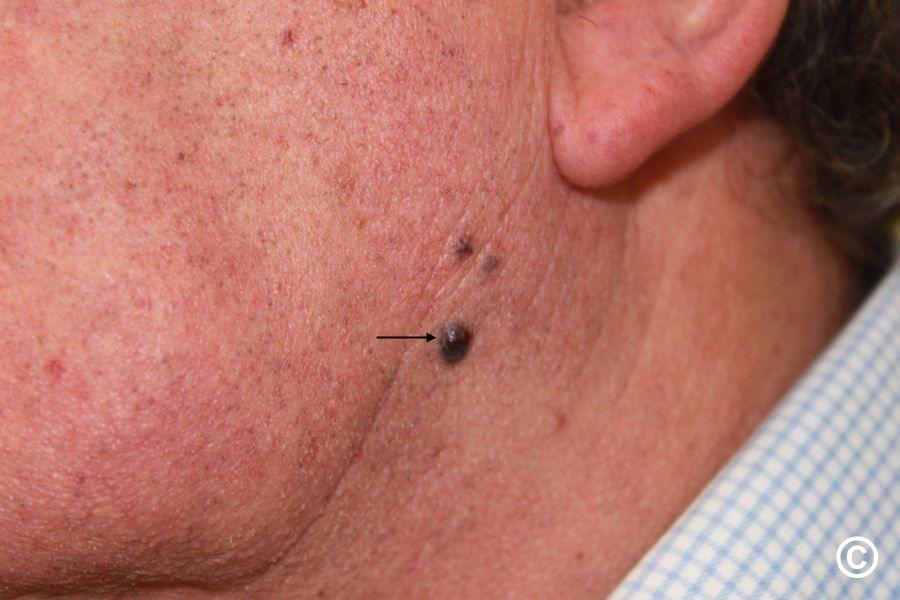
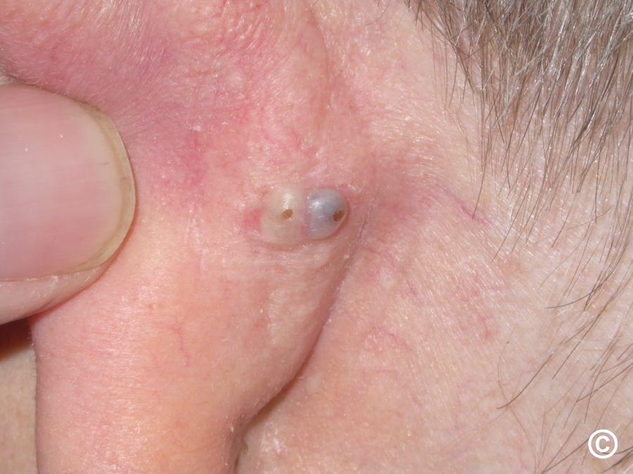
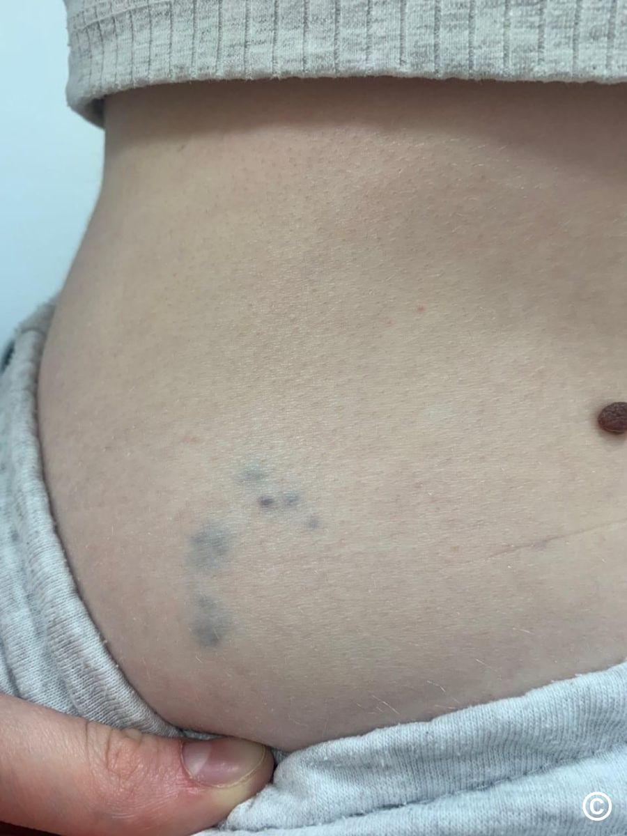
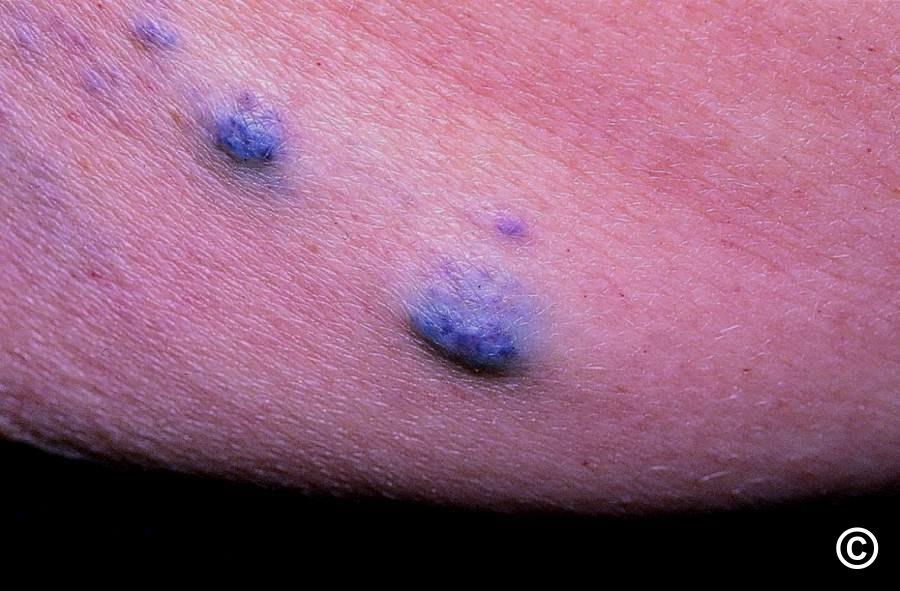
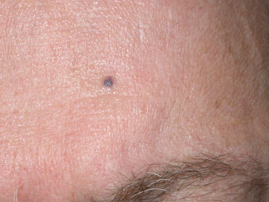
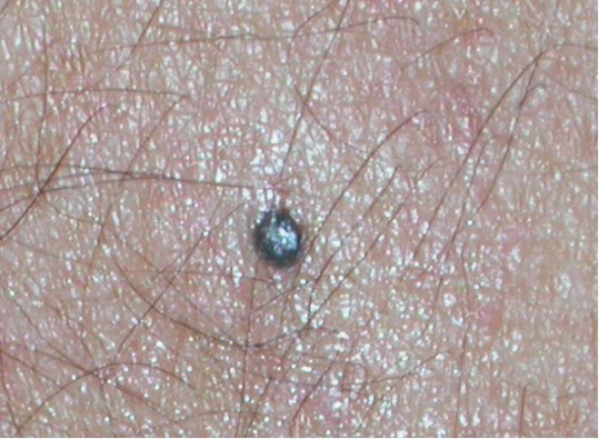
  • Usually present as a small, regular, blue or blue-grey smooth papule or macule less than 1cm in diameter
  • A cellular blue naevus is a variant of a blue naevus. Clinically lesions are usually 1-3 cm and found most commonly on the buttocks and sacral regions, and occasionally on the hands and feet

Dermoscopic features (hover over terminologies for description)

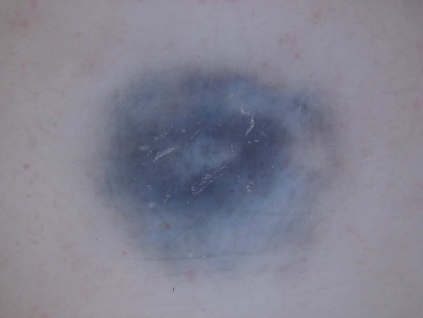
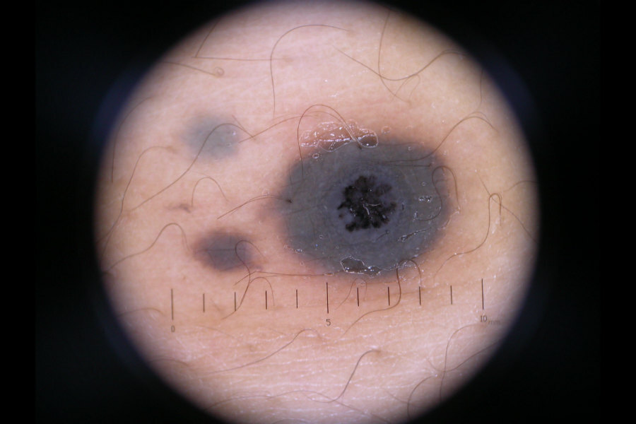
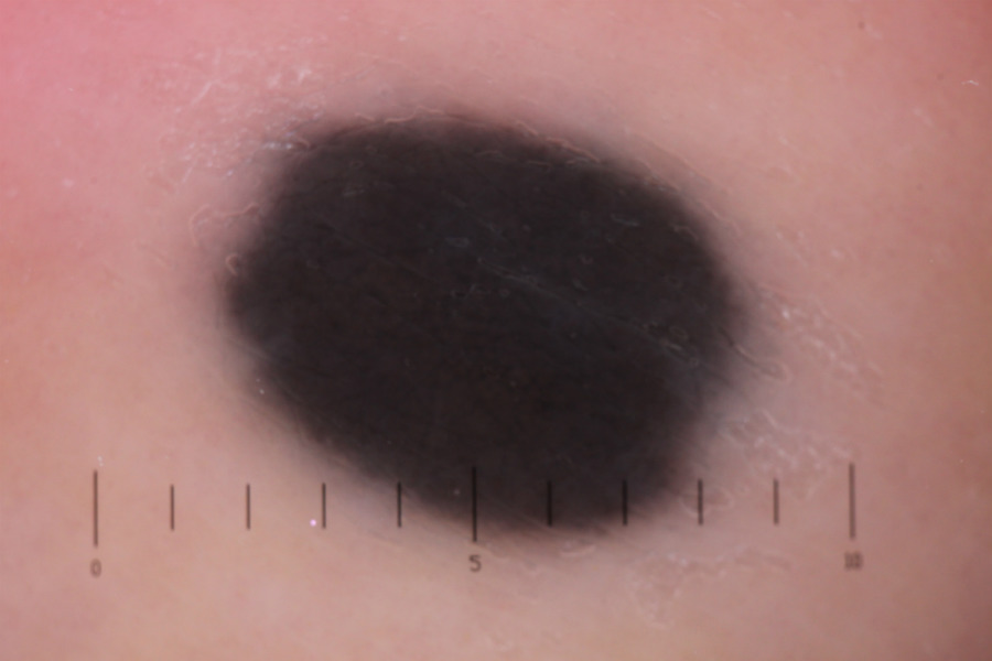
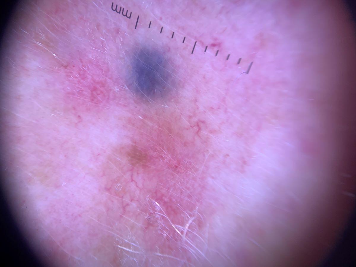
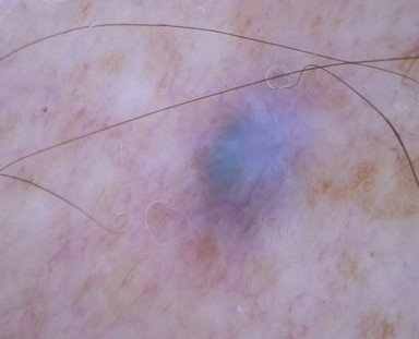
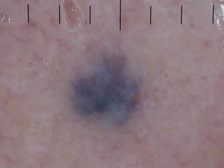
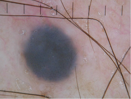
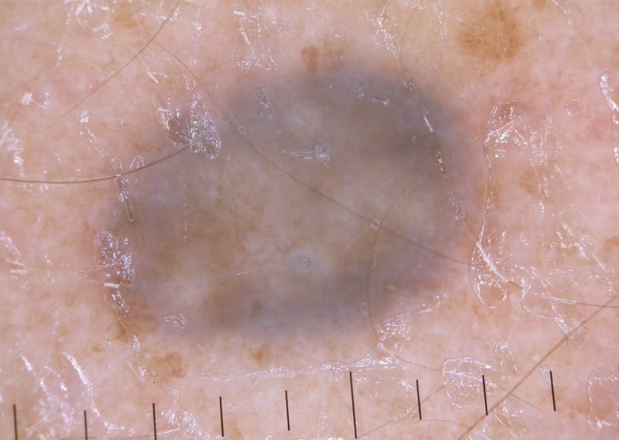
  • Homogenous blue-grey pigmentation, sometimes with white areas
  • Devoid of structures i.e. structureless 

Differential diagnosis - benign

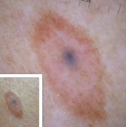
  • Combined melanocytic naevi in which melanocytes are found at different levels of skin. The most common combination is a blue naevus with a common brown acquired naevus. Typically, the multicomponent structure of combined naevi exhibit a reasonably symmetrical appearance, which is consistent with already established findings that benign naevi tend to exhibit symmetry. The structureless blue part of the lesion is most commonly central, or off-centre, usually covering about 30% of the lesion. If located centrally, the blue structureless area is surrounded by another pattern, mostly commonly a brown globular pattern or pigment network. Benign lesions should lack dermoscopic melanoma-specific features
     
  • Tattoos - these can be 'real' tattoos or iatrogenic following skin injury
     
  • Vascular lesions such as venous lakes and darker angiomas
     
  • Giant comedones
     
  • Glomulovenous malformations and the Blue Rubber Bled Naevus syndrome. Both are rare, the former tend to be painless unless there is an extreme change in temperature, the latter are characteristically painful (especially at night)

Red flags for possible melanoma

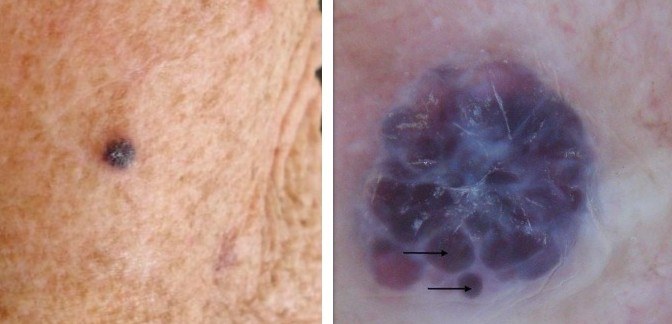
  • Combined naevi can take on a similar appearance to melanoma, the latter should be considered if the blue area is greater than 30% or extends to the edge of the lesion, if blue is combined with a colour other than brown, in chaotic lesions (asymmetry of structures or colour), and lesions with any dermoscopic melanoma-specific features
     
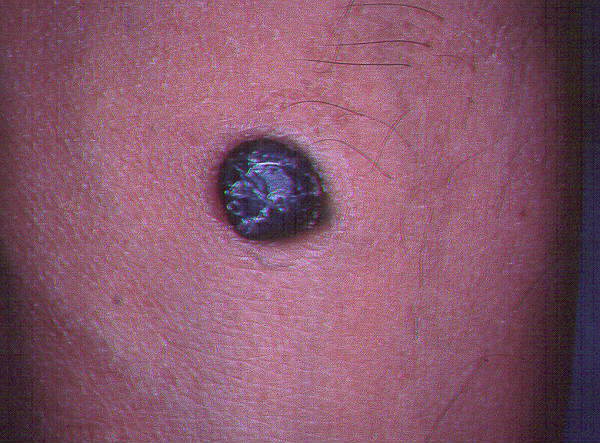
  • Melanoma can very occasionally present as a firm blue nodule, larger than one would expect for a blue naevus and showing signs of sustained growth. One or more firm blue nodules could be a presentation of metastatic melanoma
     
  • Transformation to a malignant blue naevus is exceedingly rare, but has been reported, predominantly with cellular blue naevi. While blue naevi should not be excised prophylactically, significant change to a long-standing lesion needs to be treated with caution

Clinical Images

Please refer to notes on image rights at bottom of the page with regards to individual image ownership.
Management

  • Blue naevi are harmless and do not need to be excised
  • If there is uncertainty about the diagnosis the patient should be referred to Secondary Care on an urgent suspected cancer pathway  

Disclaimer - the author PCDS cannot accept responsibility for any misleading or incorrect statements, and the management of individual patients remains the direct responsibility of the individual doctor. We do however hope that visitors to this site can contact us regarding comments that are considered misleading or incorrect so that we can continue to improve the site.

Image Rights - The PCDS would like to thank Dermatoweb, DermQuest (Galderma), and others who have contributed images. All named individuals and organisations maintain copyright for the relevant images.

Quick Links

The following pharmaceutical companies have had no involvement in the content of this website or in our conference programmes


Almirall
Galderma
Glenmark
Johnson & Johnson
La Roche-Posay
LEO Pharma
Pierre Fabre
Schuco