Basal cell carcinoma: solid (syn. nodular / cystic)

LAST UPDATED: Jun 05, 2026

Patient Information Leaflet
Link: Basal cell carcinoma

https://www.pcds.org.uk/patient-info-leaflets/basal-cell-carcinoma
Introduction

The solid basal cell carcinoma (BCC), sometimes referred to as nodular or cystic, is the most common type of basal cell carcinoma.

This chapter is set out as follows:


Aetiology

For more information refer to the related chapter Basal cell carcinoma - an overview.


Clinical findings

Distribution

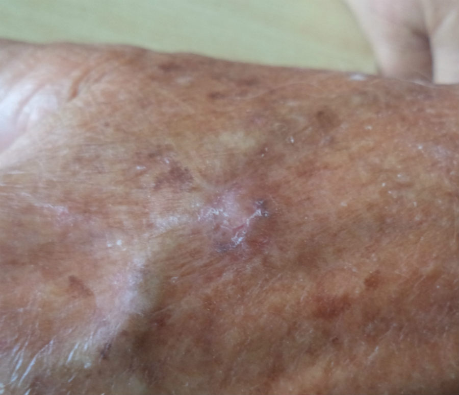
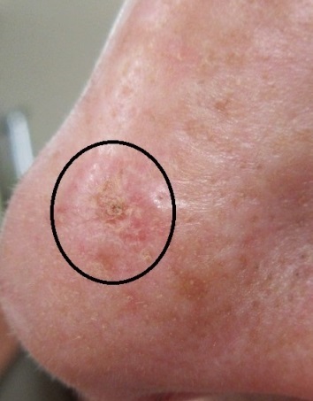
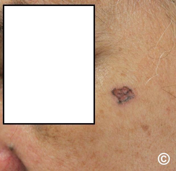
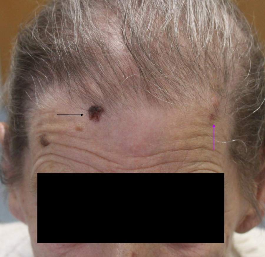
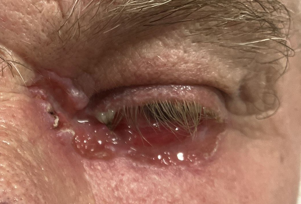
  • Any site can be involved; the head and neck are the most commonly affected
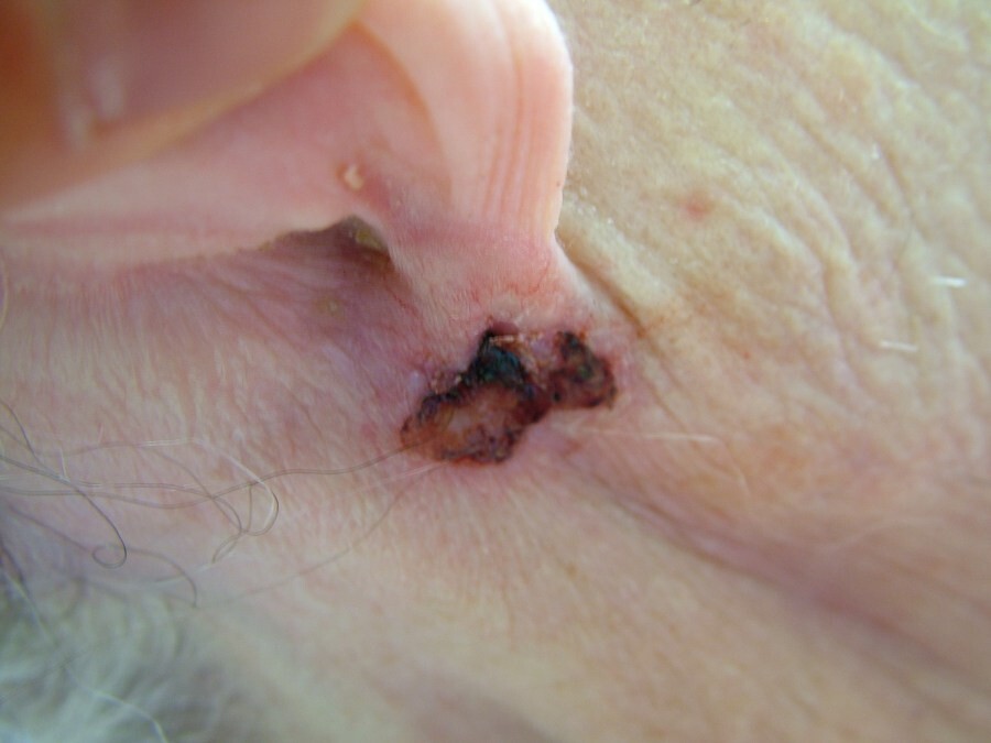
  • The medial canthus and behind the ears are not uncommon sites; a thorough skin examination is required 

Morphology

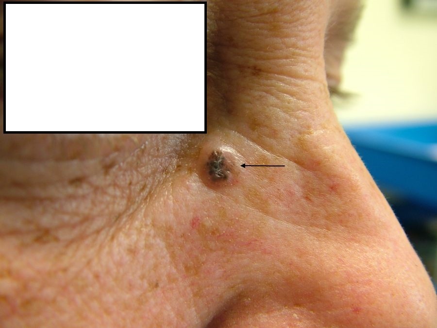
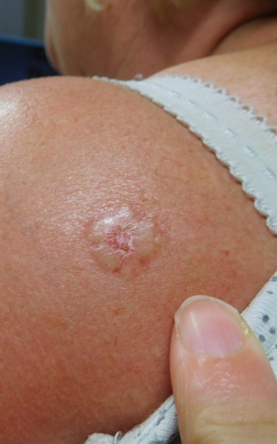
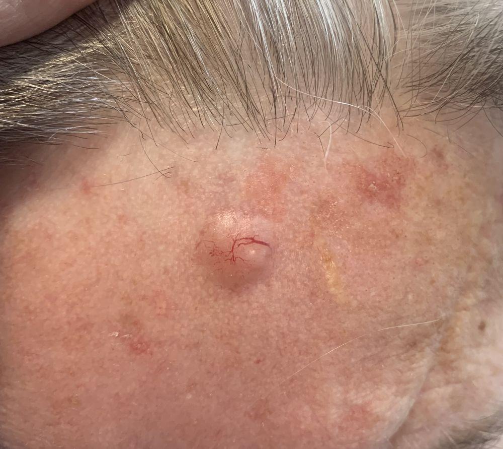
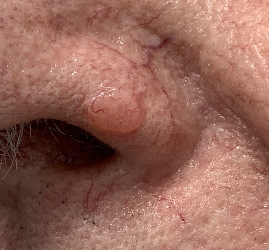
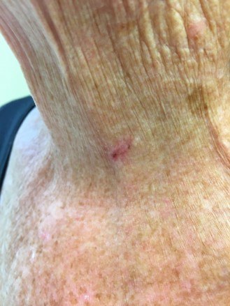
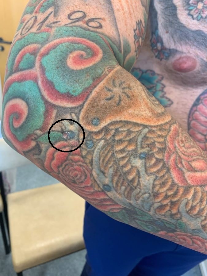
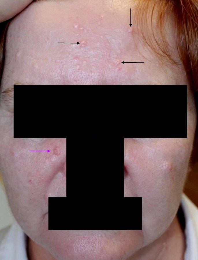
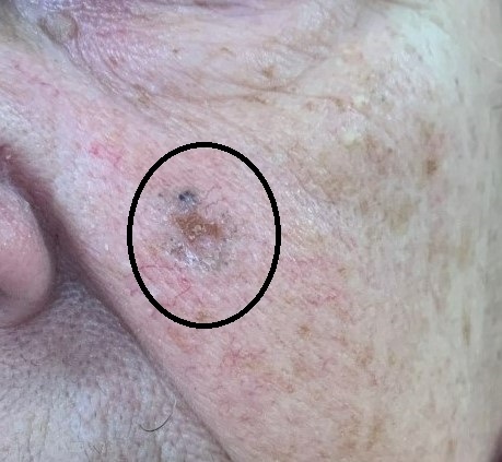
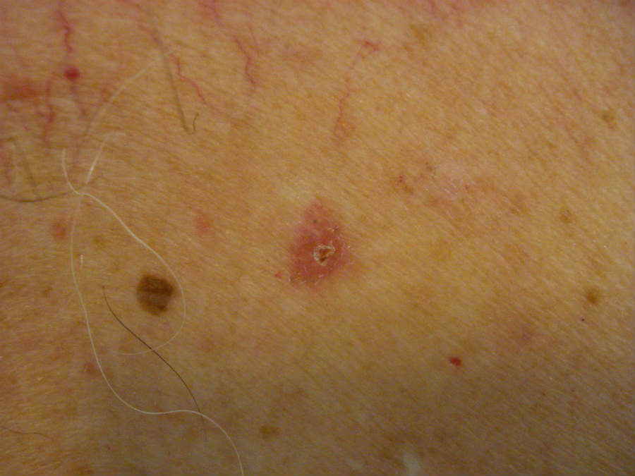
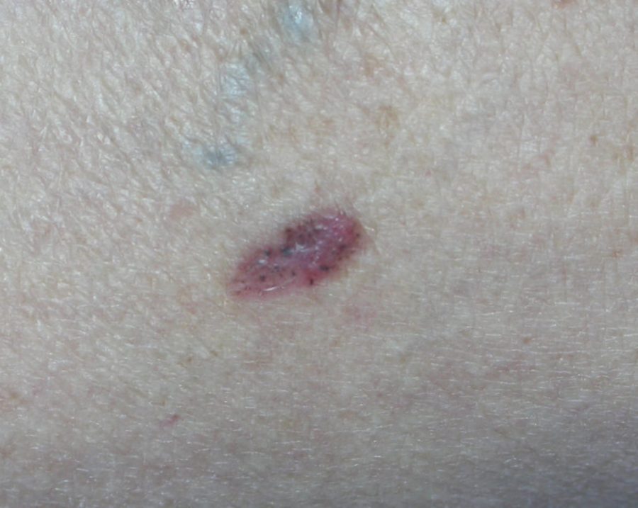
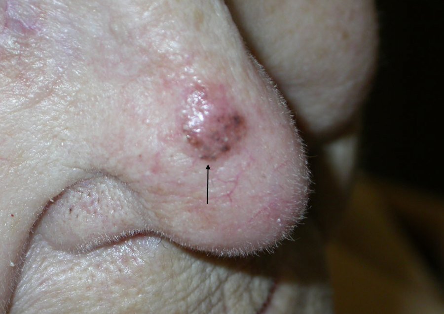
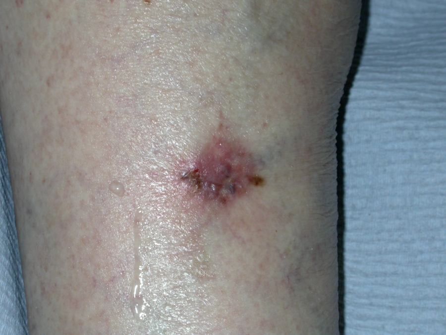
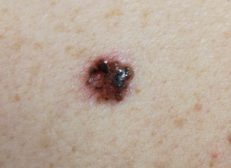
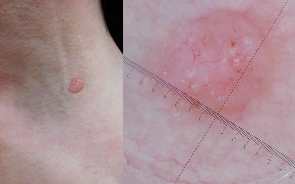
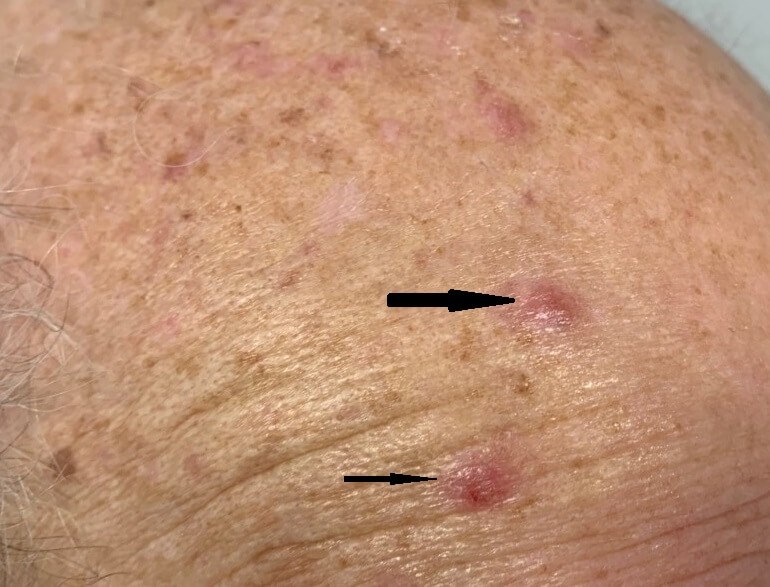
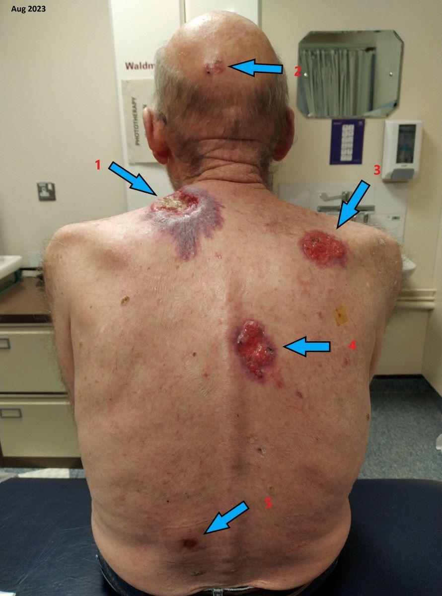
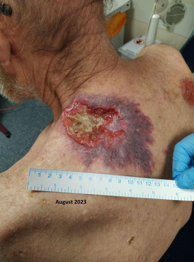
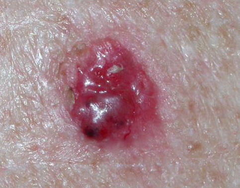
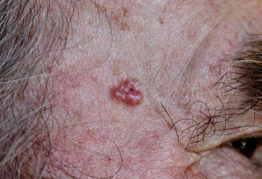
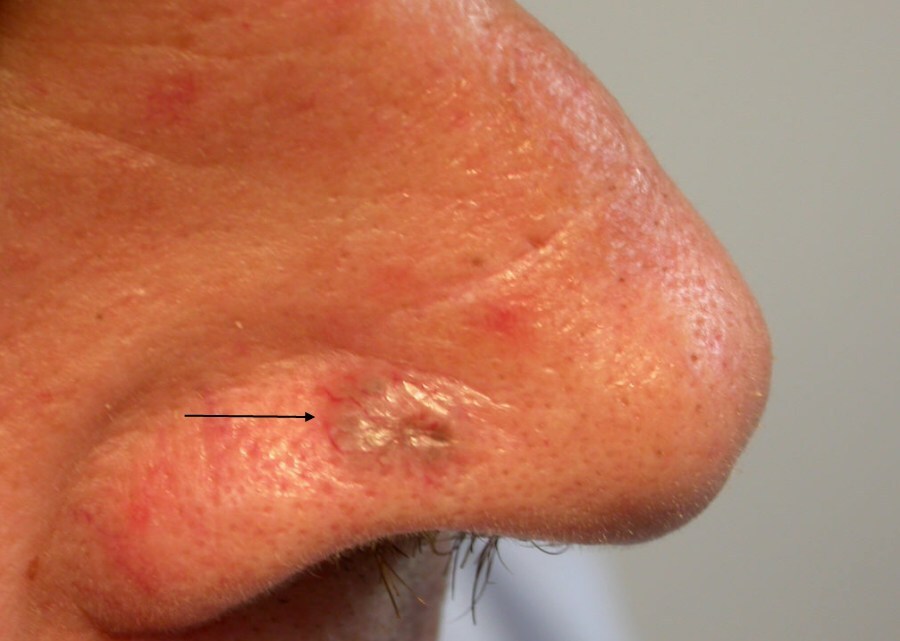
  • Commonly raised; can be flat
  • Shiny / translucent, rolled edge, telangiectasia (more peripheral)
  • With time the nodule becomes more irregular and ulcerates
  • Very firm

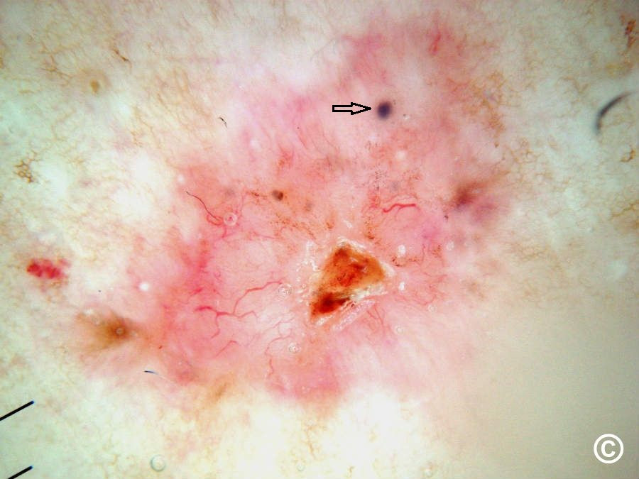
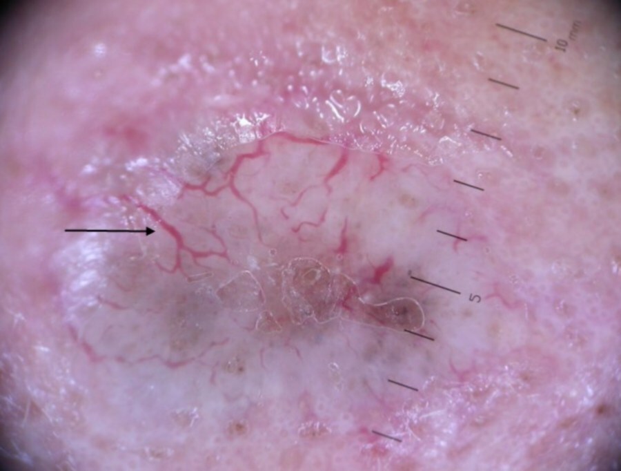
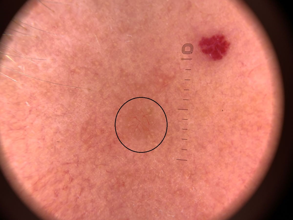
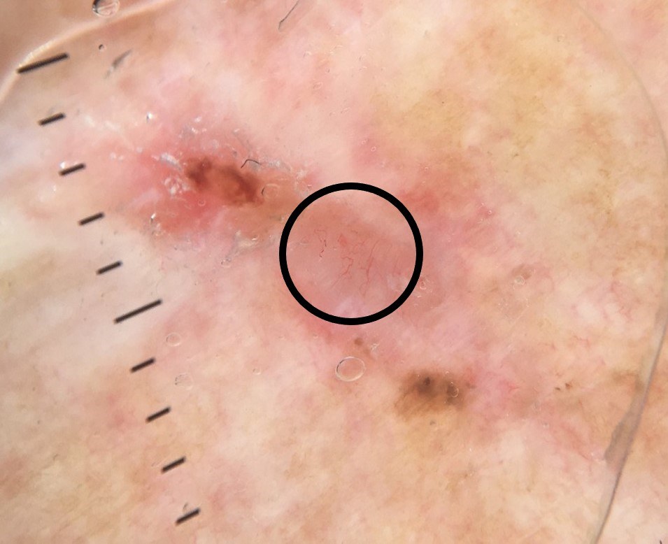
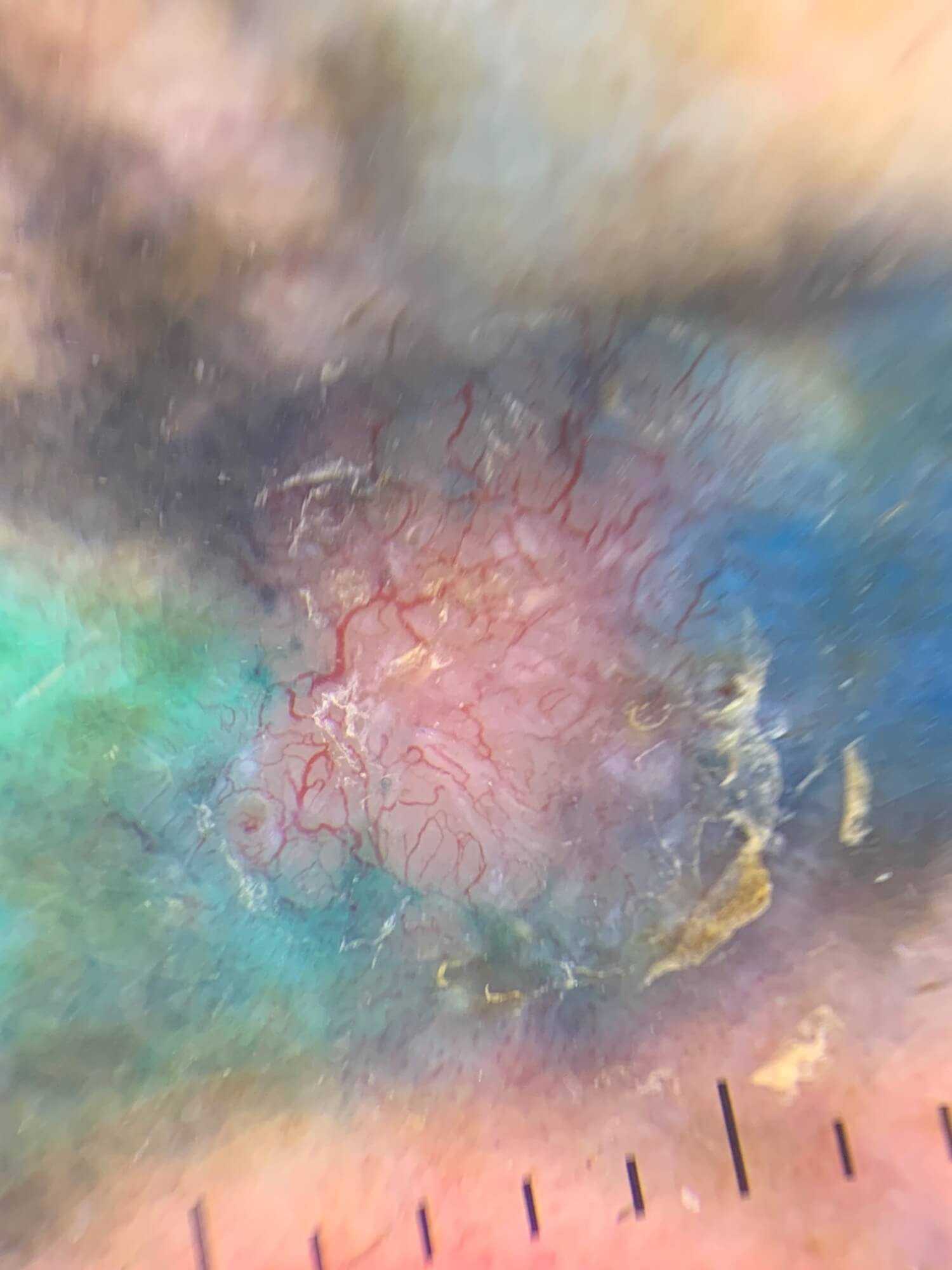
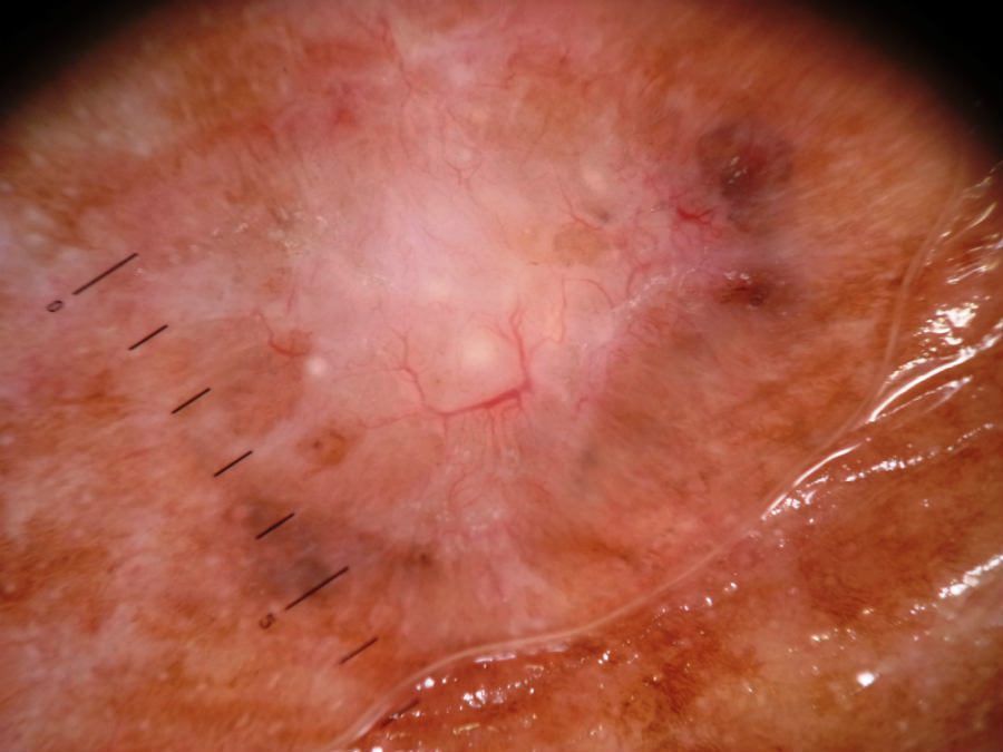
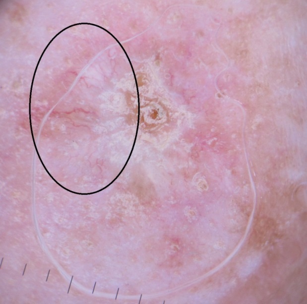
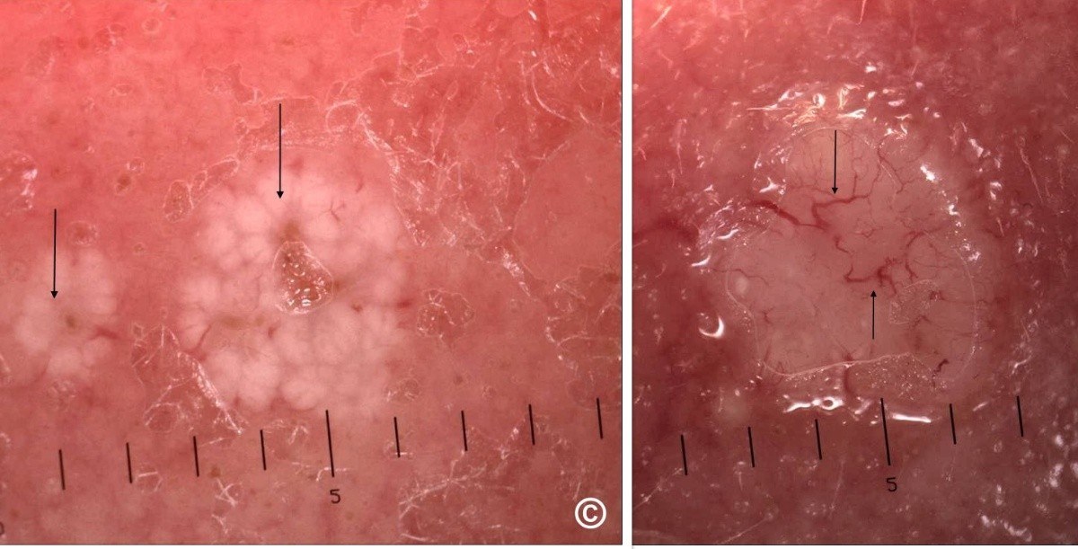
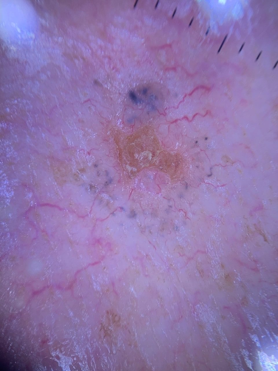
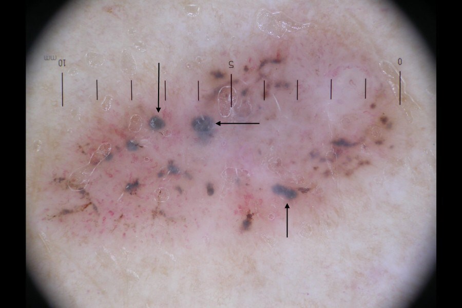
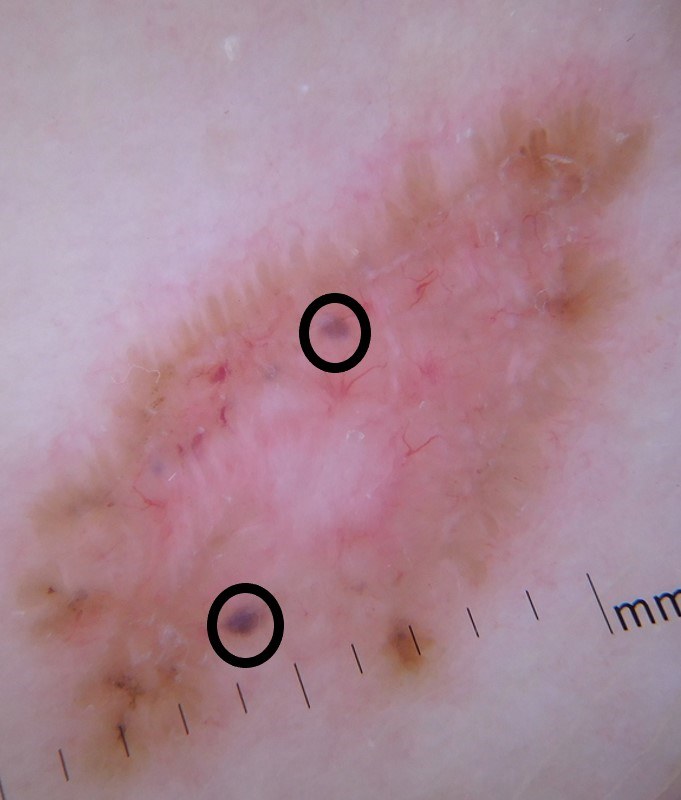
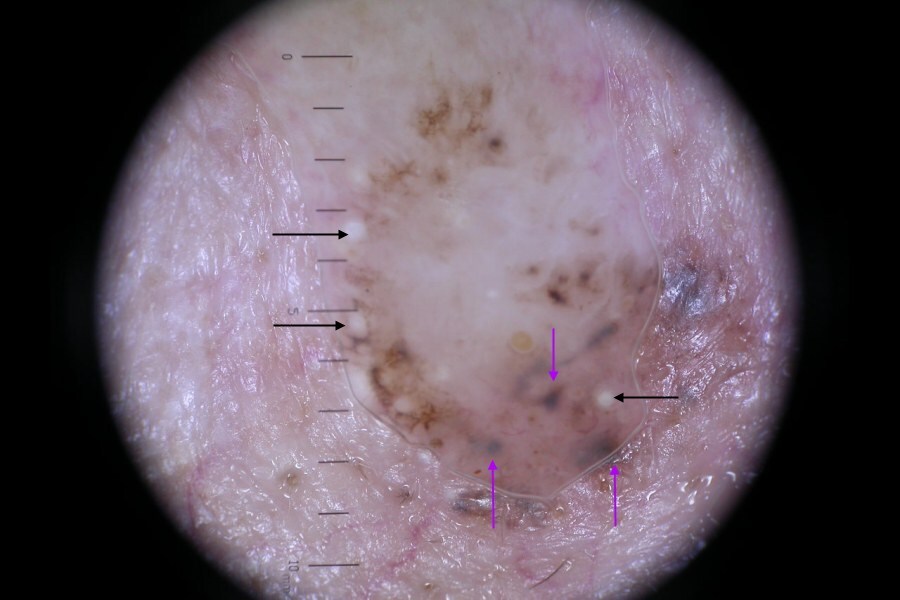
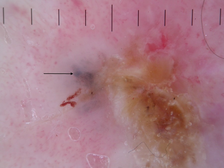
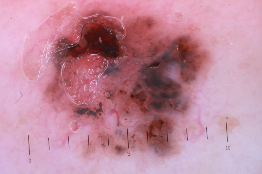
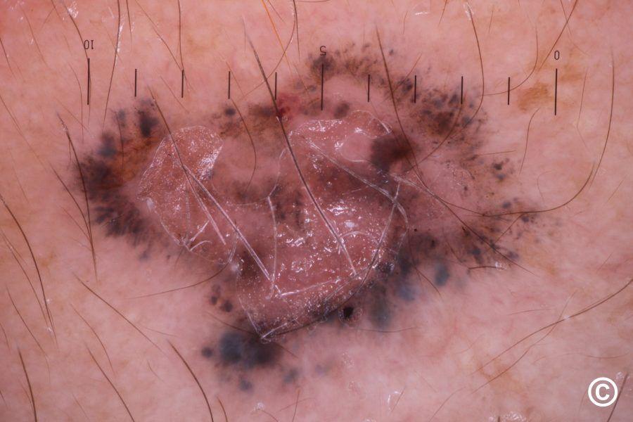
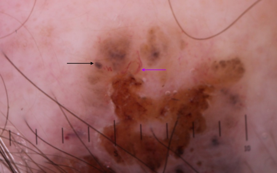
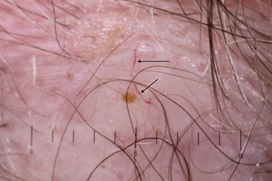
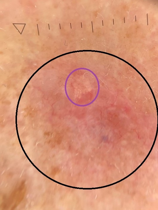
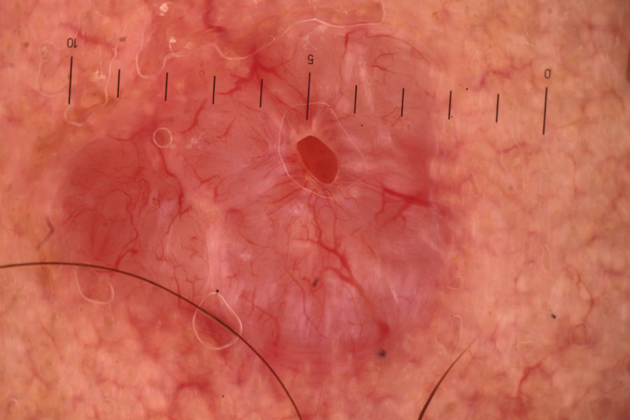
Dermoscopic features (hover over terminologies for description)

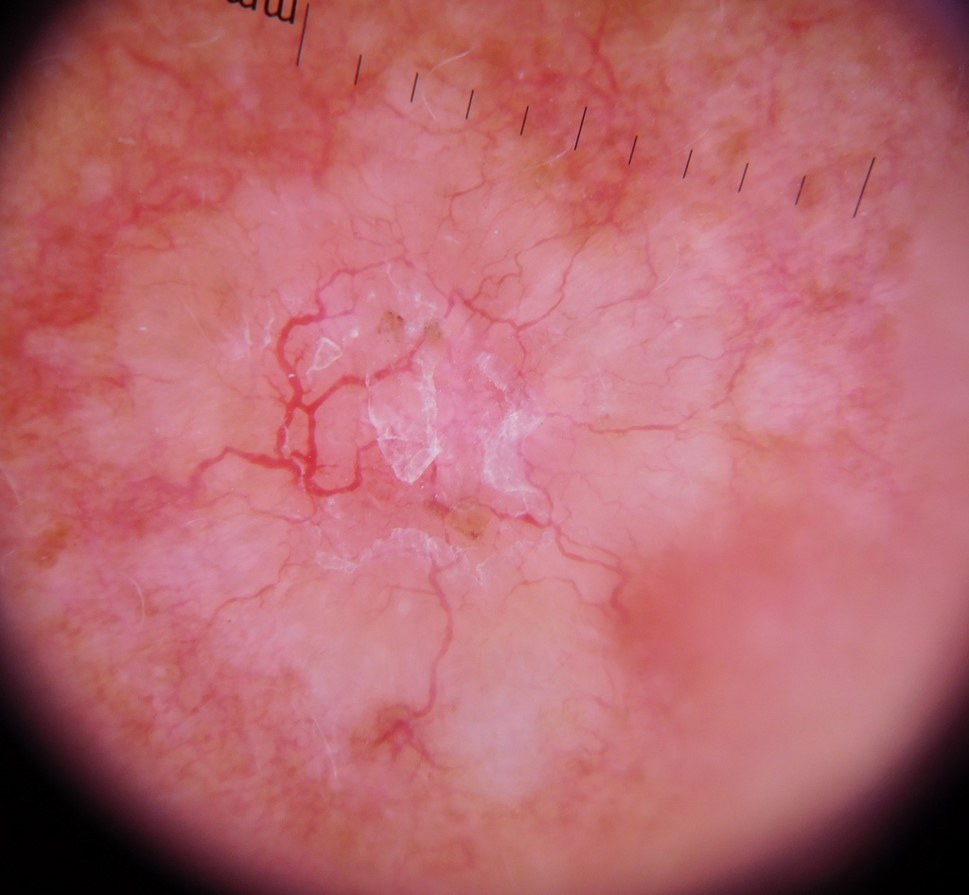
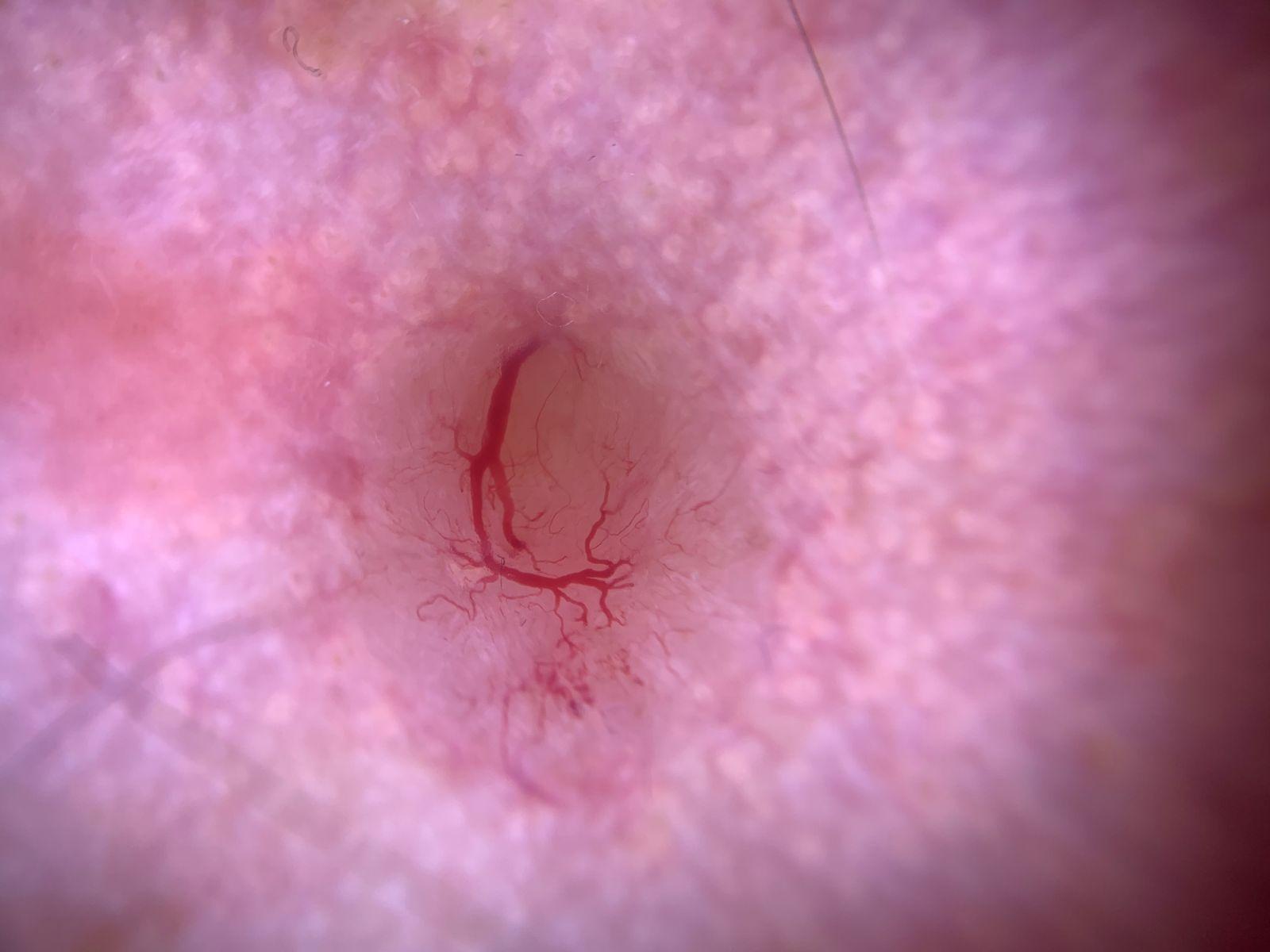
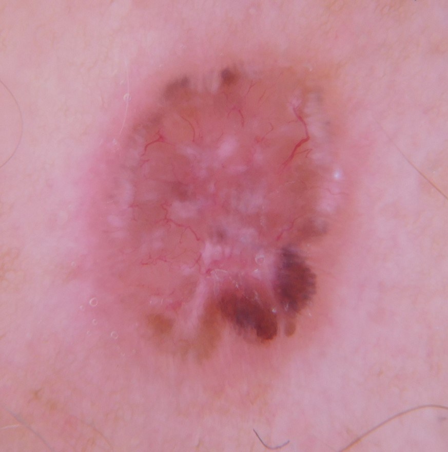
  • Pinky white translucent or gelatinous background
     
  • Sharply focused arborising (branching) vessels (for more information on vessels refer to the chapter Vessel patterns in dermoscopy)
     
  • Ulcerations or erosions
     
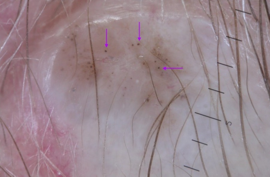
  • Blue / grey clods (sometimes referred to as ovoid nests) or dots. Never a pigment network
     
  • Shiny white structures - strands (streaks) or blotches, seen primarily in the polarised mode
     
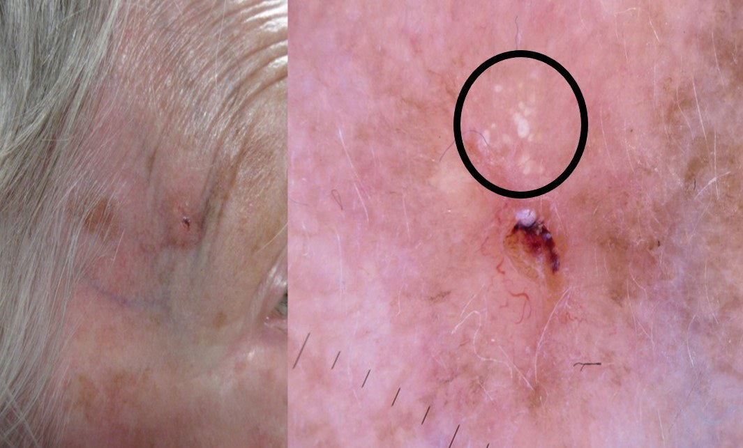
  • MAY globules - multiple aggregated yellow-white globules (clods). These differ from milia-like cysts as they are grouped and observed equally with non-polarised and polarised light (milia-like cysts are best seen with non-polarised light). MAY globules are positively associated with deeper-seated and histologically higher-grade tumour subtypes (eg micronodular), and negatively associated with superficial BCC

BCC in skin of colour

  • The diagnosis of BCC in skin of colour can be difficult due to its rarity and pigmentation
  • More than 50% of such cases are pigmented with a ‘pearly’ brown or black appearance and a rolled border
  • Nodular BCC are the predominant subtype; infiltrative BCC are uncommon
  • In a study of 66 BCC in skin phototypes IV or V the most common dermoscopic features were pigmented peripheral clods (61.7%), blue-white structureless areas (53.4%), ulceration (48.4%), and short arborising vessels (46.7%)

Clinical Images

Please refer to notes on image rights at bottom of the page with regards to individual image ownership.
Management


Disclaimer - the author PCDS cannot accept responsibility for any misleading or incorrect statements, and the management of individual patients remains the direct responsibility of the individual doctor. We do however hope that visitors to this site can contact us regarding comments that are considered misleading or incorrect so that we can continue to improve the site.
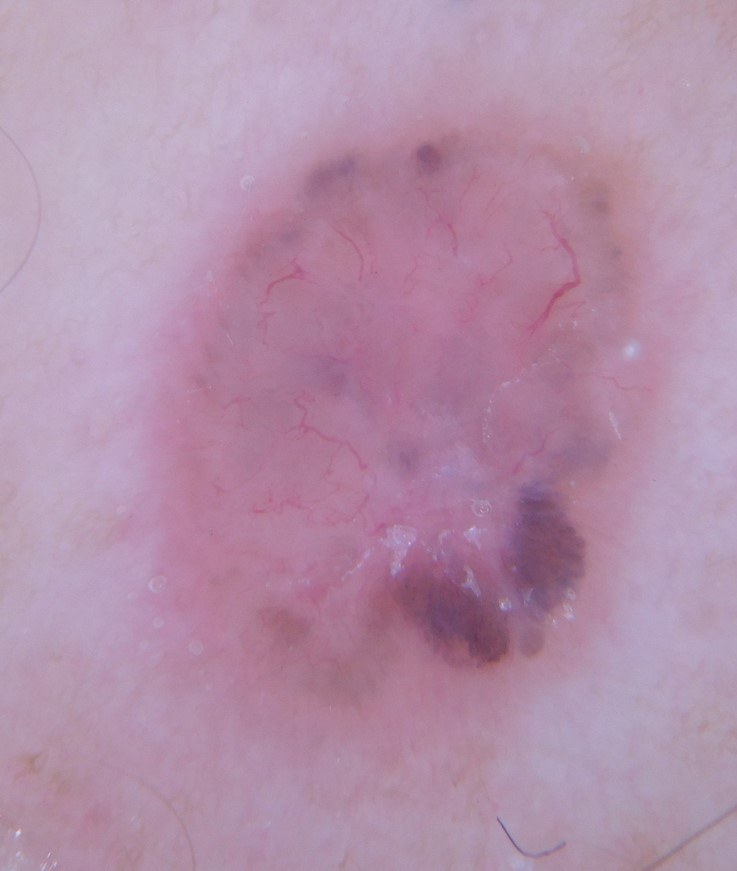
Image Rights - The PCDS would like to thank Dermatoweb, DermQuest (Galderma), and others who have contributed images. All named individuals and organisations maintain copyright for the relevant images.

Quick Links

The following pharmaceutical companies have had no involvement in the content of this website or in our conference programmes


Almirall
Galderma
Glenmark
Johnson & Johnson
La Roche-Posay
LEO Pharma
Pierre Fabre
Schuco