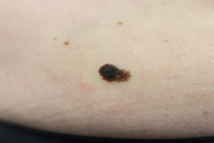
Seborrhoeic keratosis (syn. seborrhoeic wart, basal cell papilloma)
LAST UPDATED: Jun 05, 2026
Introduction
A seborrhoeic keratosis (sebK) is a benign (harmless) overgrowth of epidermal keratinocytes of unknown aetiology that is prevalent in middle-aged and older individuals.
This chapter is set out as follows:
History
- Age of presentation - most commonly present from the fourth decade onwards; younger adults can be affected
- Symptoms
- Can be asymptomatic or itch
- May grow
- Non-tender unless traumatised
- Part / all of the lesion can come away with minimal trauma
Clinical findings
Clinical examination
- Distribution
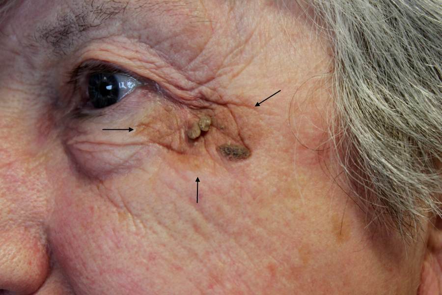
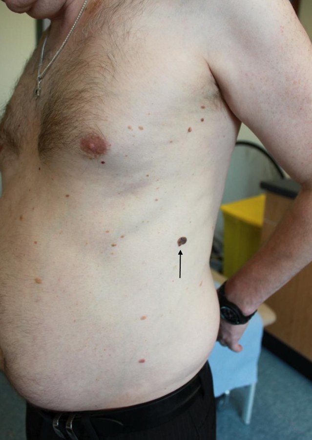
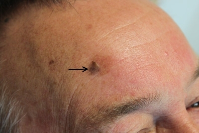
- The trunk and face are commonly affected; can arise on almost any body site
- It is not uncommon to find multiple lesions; some patients have very large numbers (especially if there is a family history)
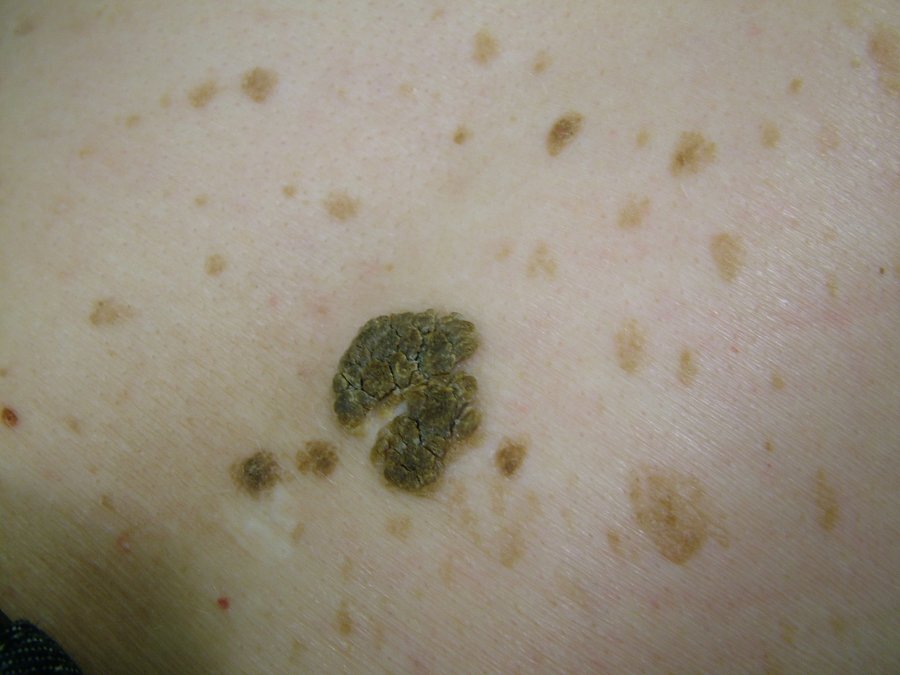
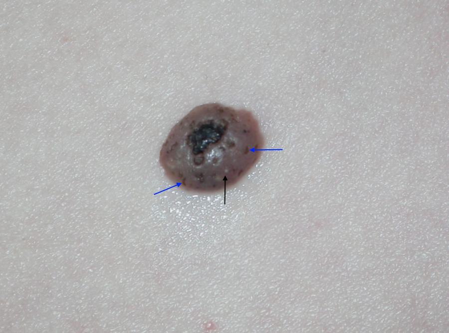
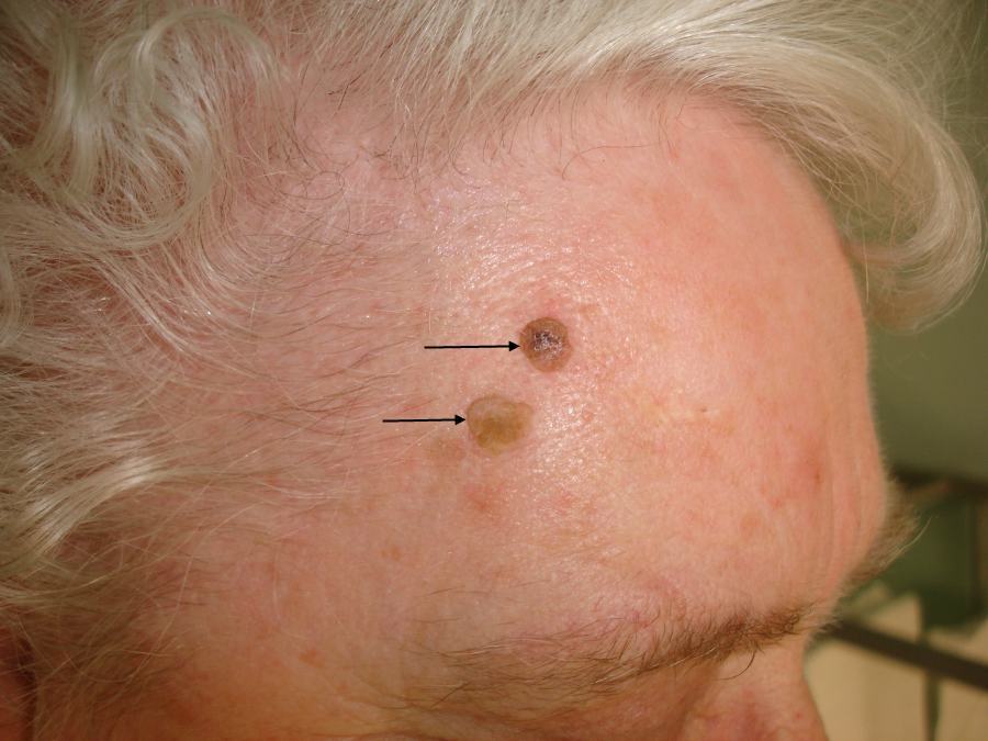
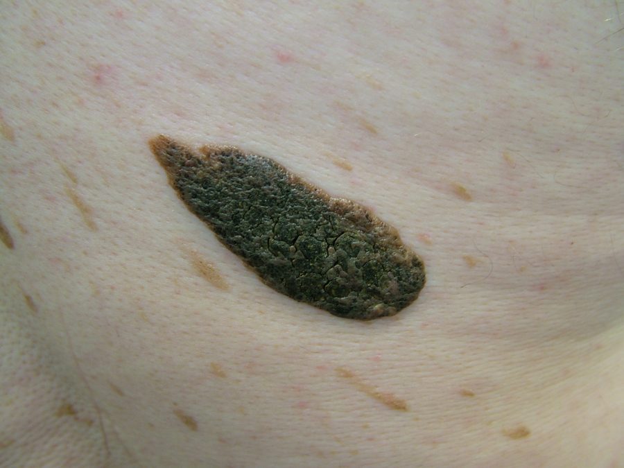
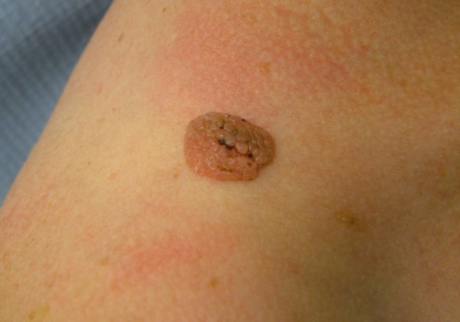
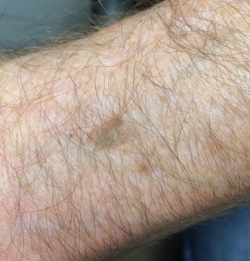
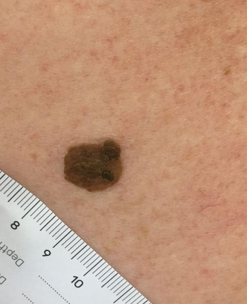
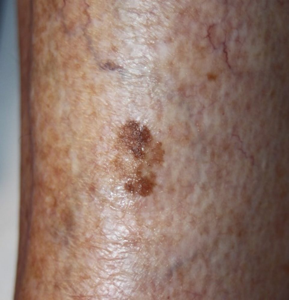
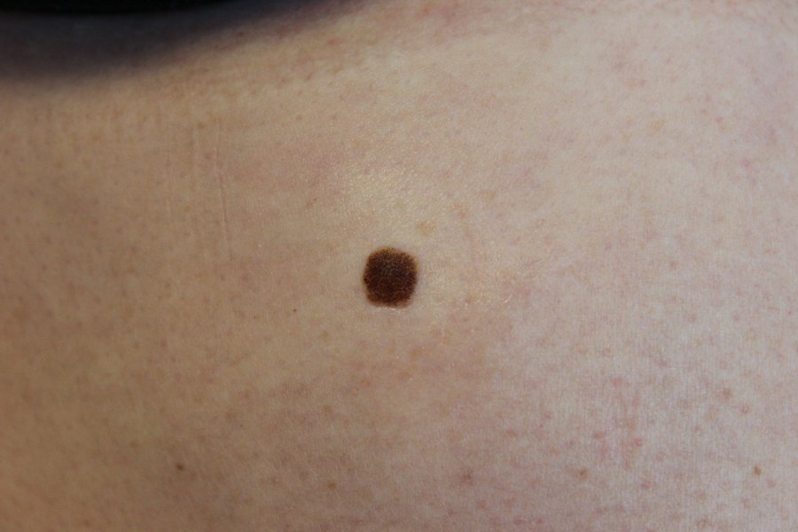
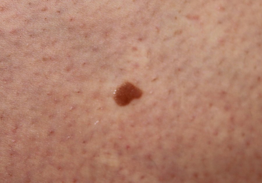
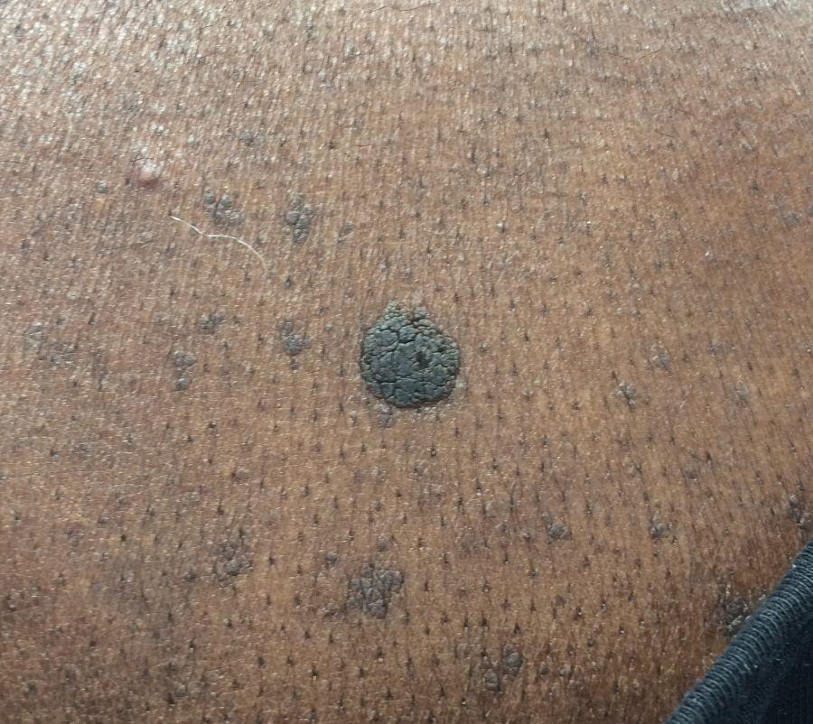
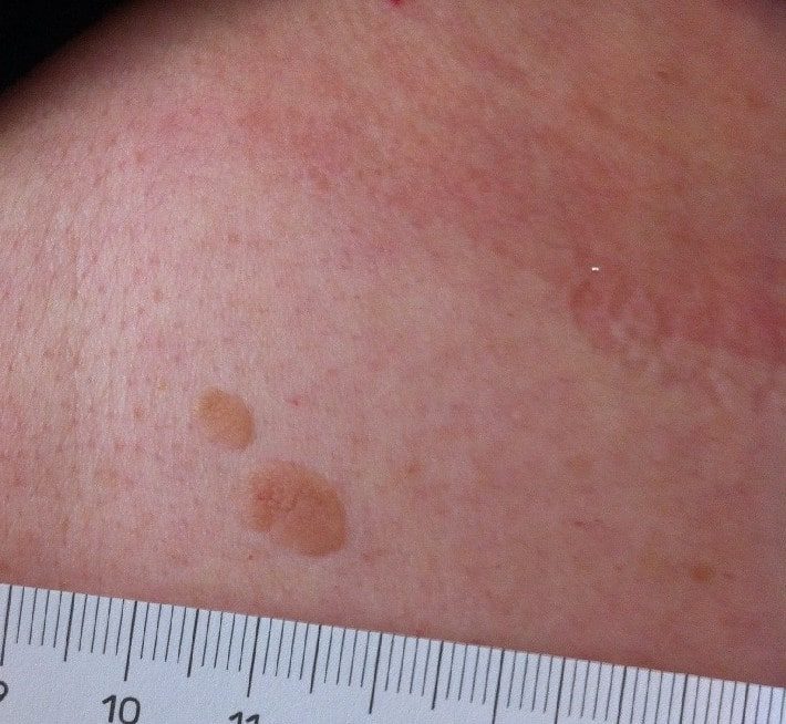
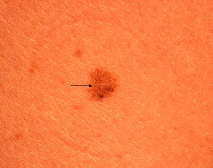
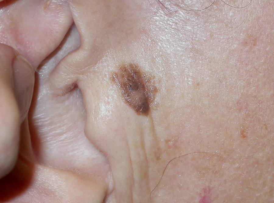
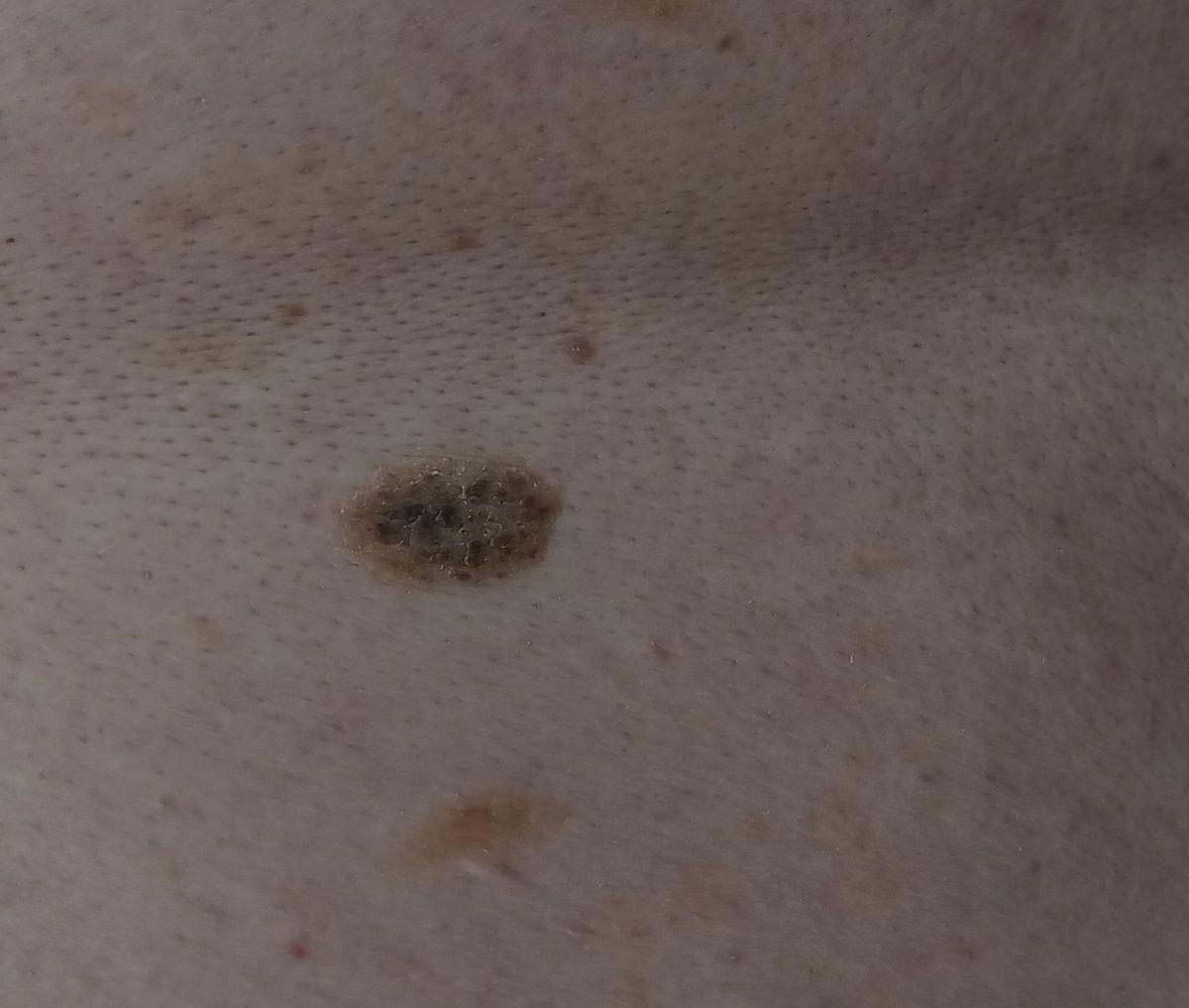
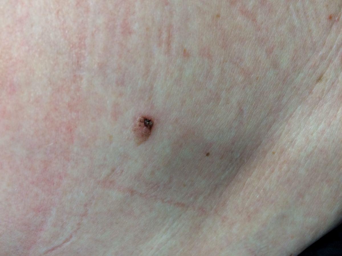
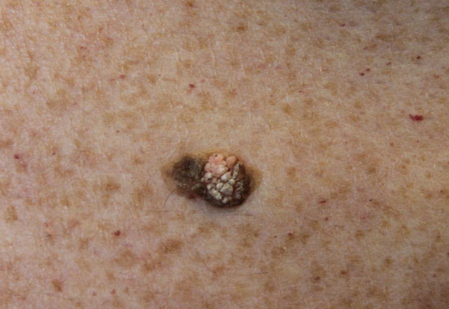
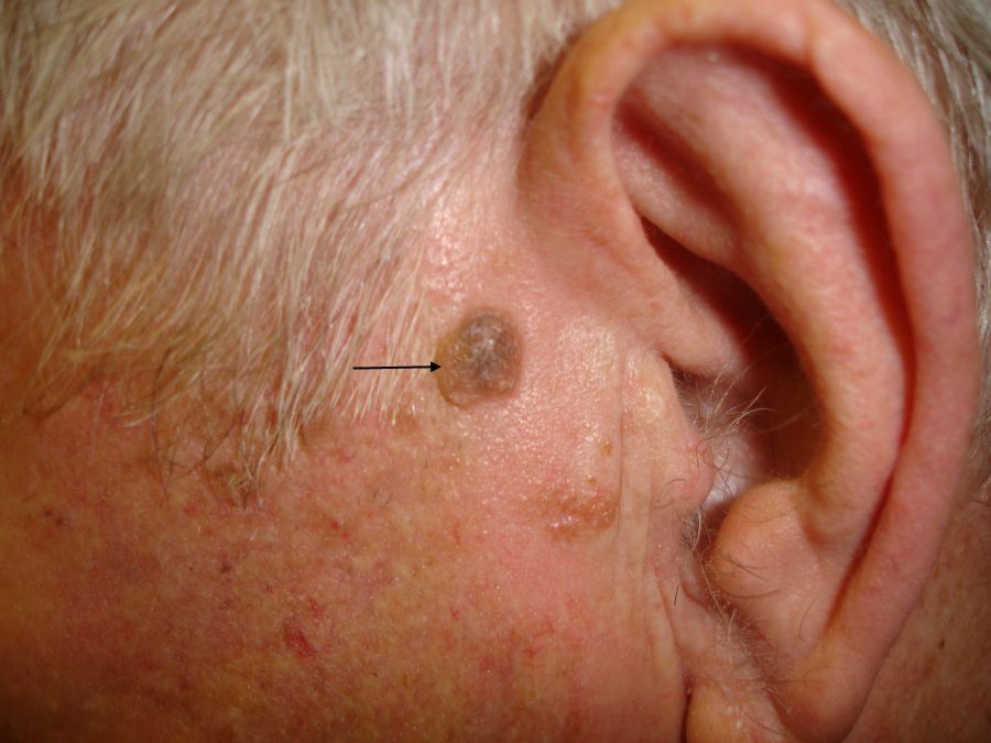
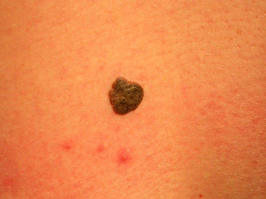
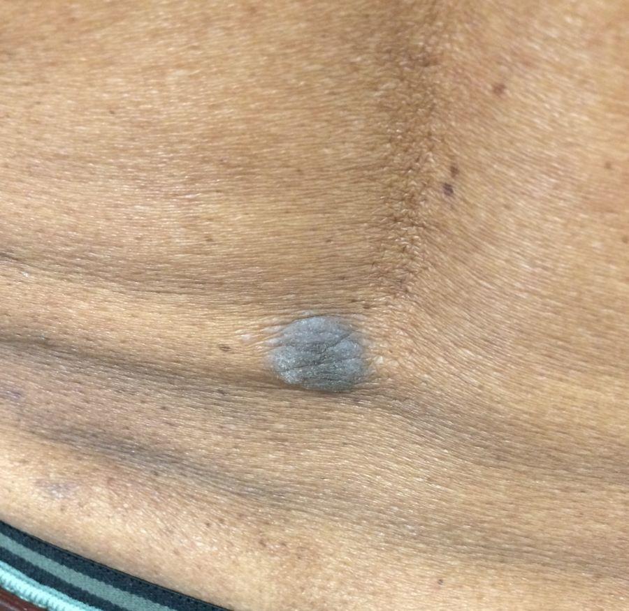
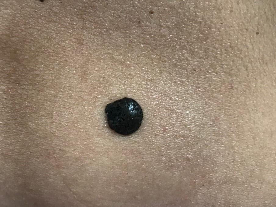
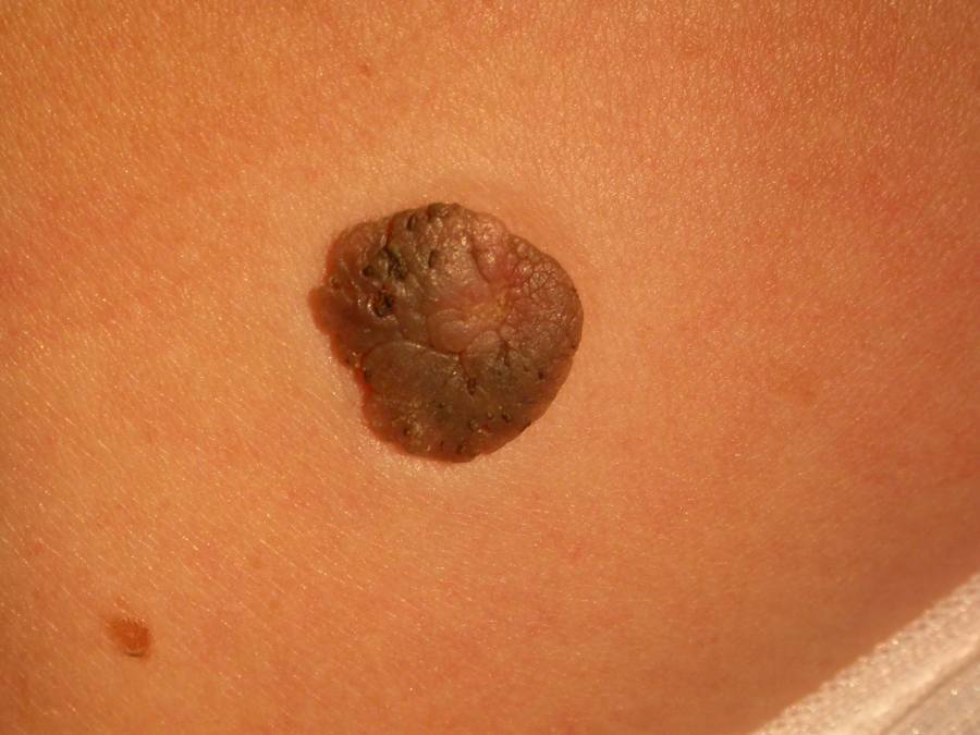
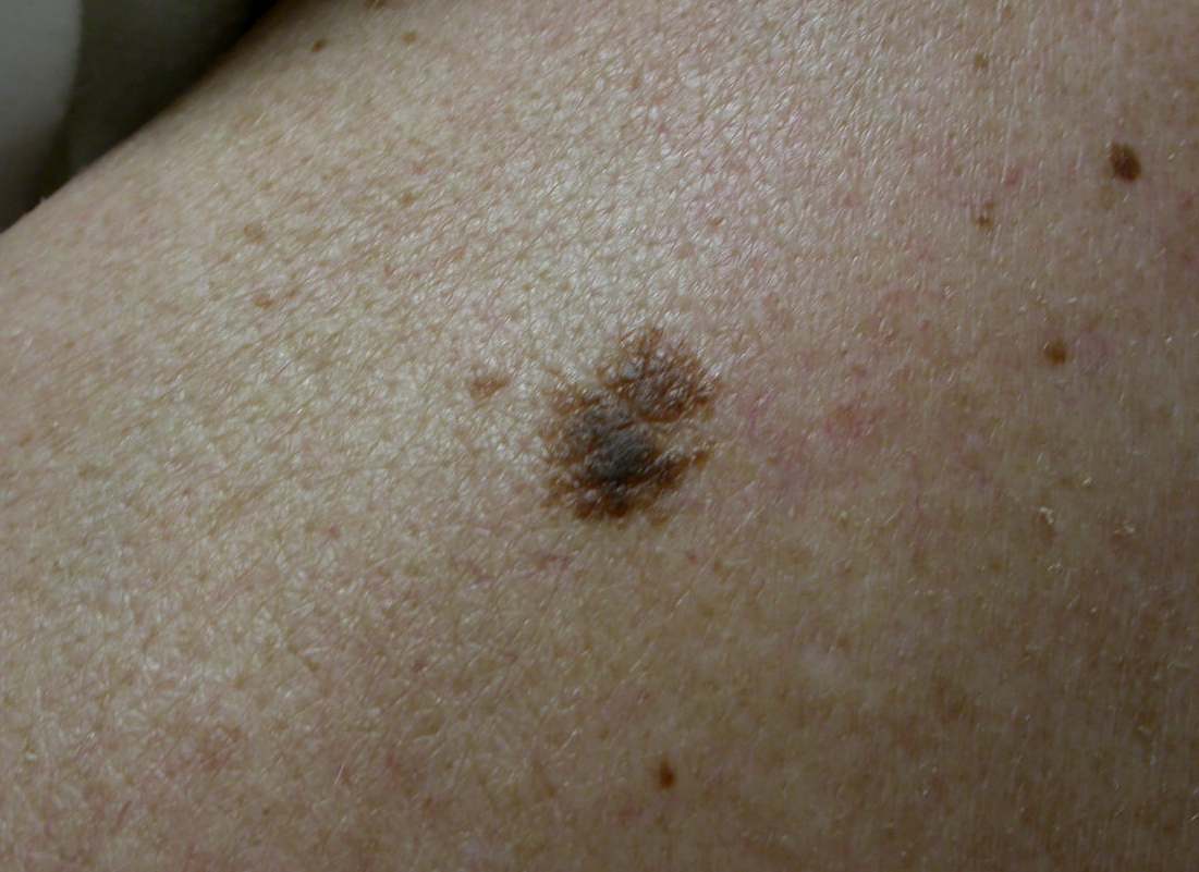
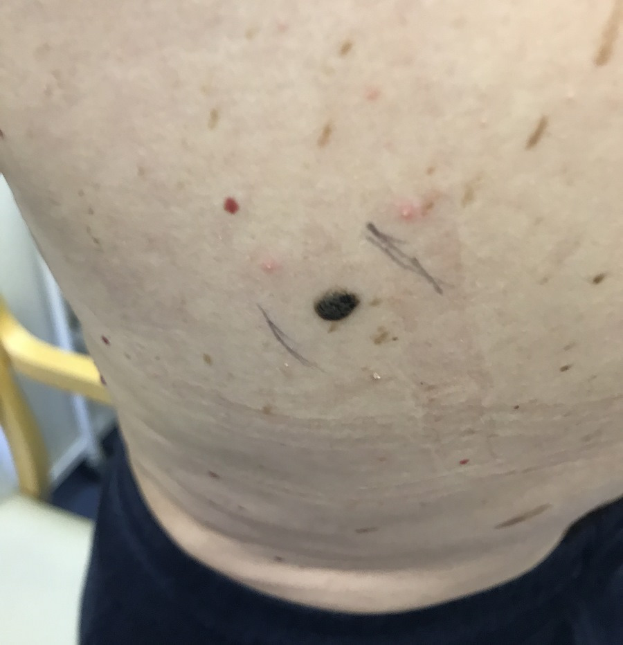
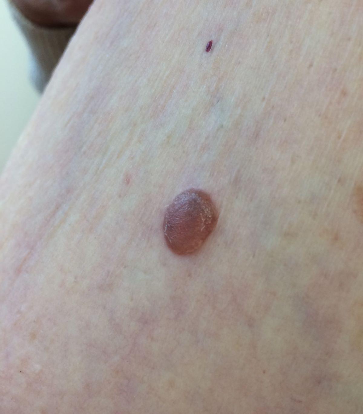
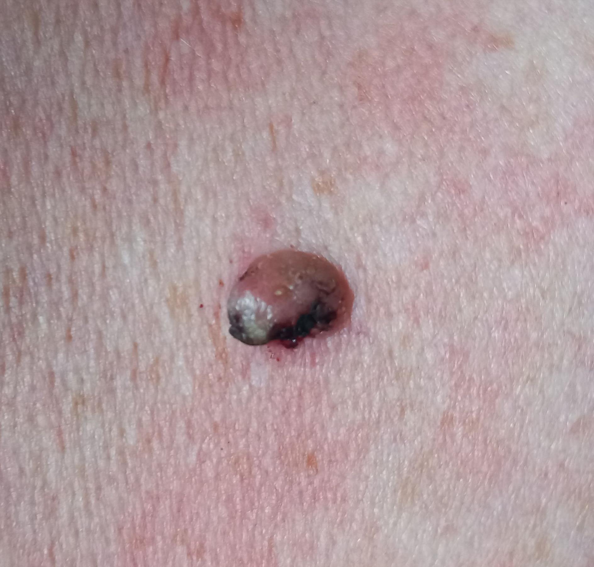
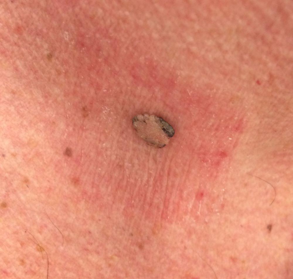
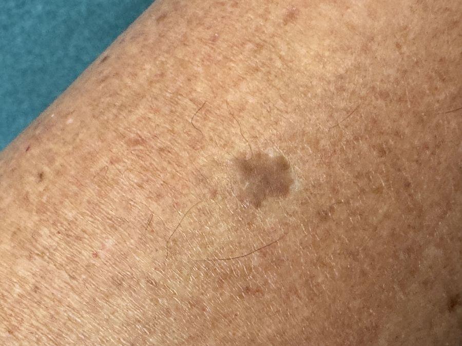
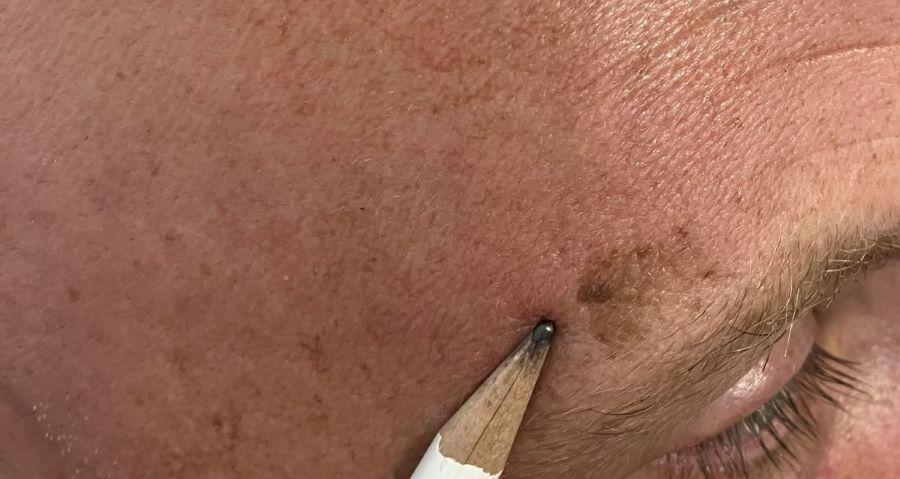
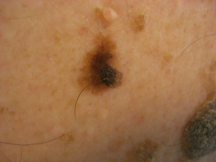
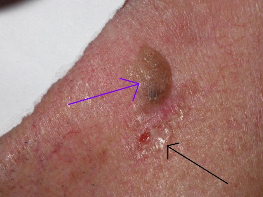
- Colour - yellow/brown to black, thinner lesions are paler, and traumatised lesions appear inflamed
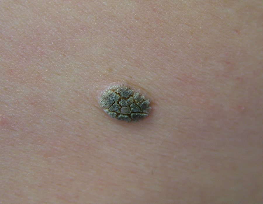
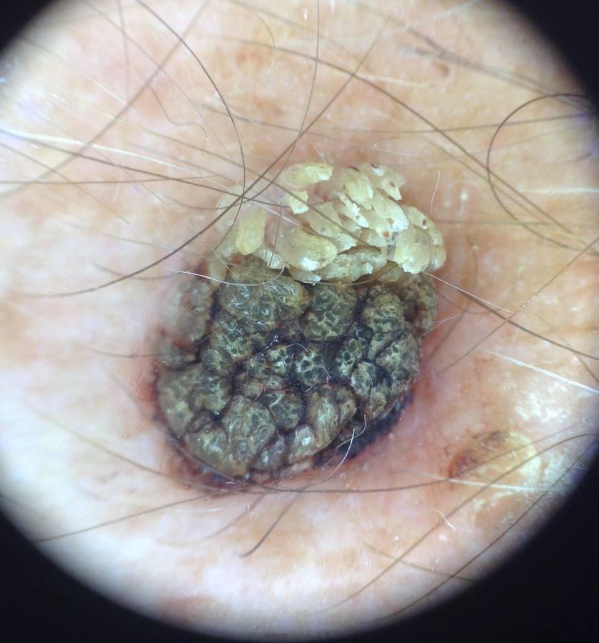
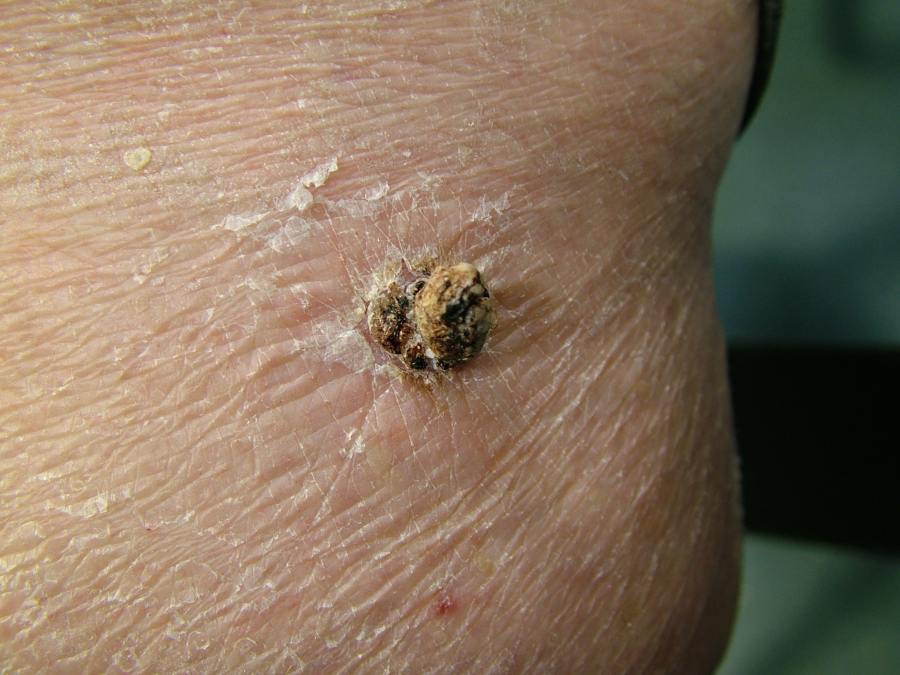
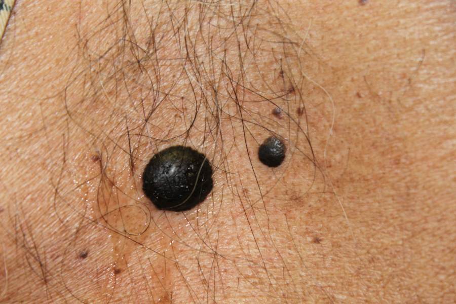
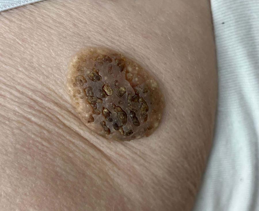
- The most common presentation is that of a thickened lesion with the following characteristics:
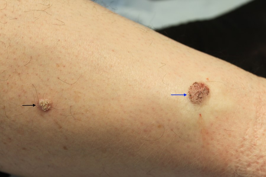
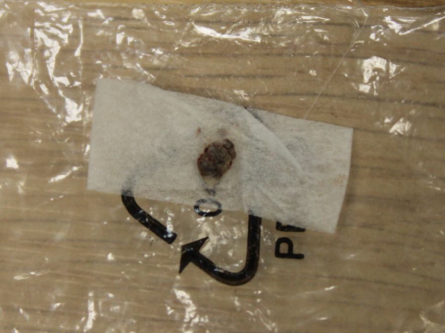
- Scaly or 'greasy' in appearance, sometimes with an irregular verrucous surface
- ‘Stuck-on’ - the lesion sits on top of normal looking skin and gives the impression that it can be easily picked off
- Most are 1-3 cm in diameter; some are very large
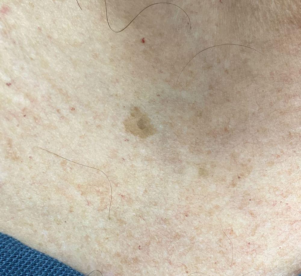
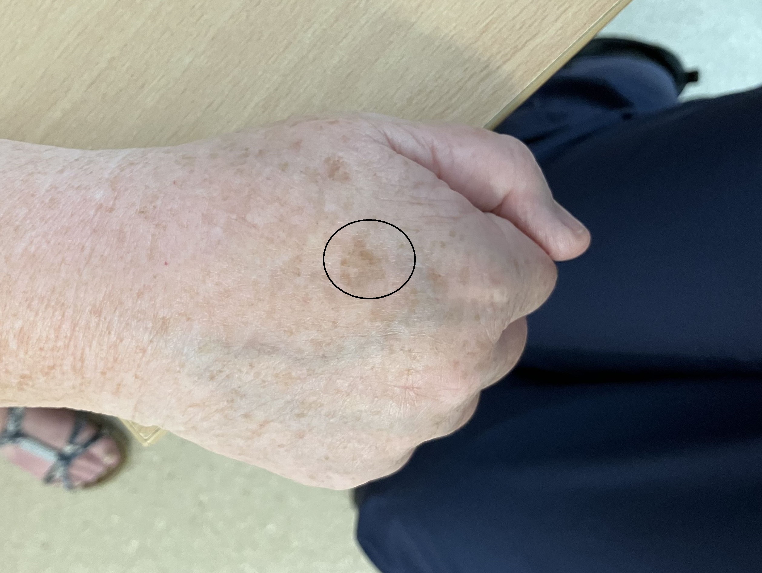
- Can grow on top of solar lentigos
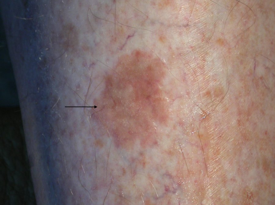
- Thinner lesions most commonly arise on areas of thinner skin (eg lower legs and forearms) and are more subtle, generally lighter in colour
- A verrucous keratosis - is where a viral wart has started to grow within a seborrhoeic keratosis. These changes are apparent both clinically and histologically
- The Leser-Trélat sign - this is characterised by the abrupt appearance of multiple seborrhoeic keratoses that rapidly increase in their size and number. There is debate as to whether or not this is a paraneoplastic phenomenon (associated with adenocarcinoma of the stomach or colon) due to the high prevalence of multiple seborrhoeic keratoses in elderly populations, however, should lesions erupt in the presence of weight loss, GI symptoms, malignant acanthosis nigricans or other 'red flags' then patients should be referred urgently for further investigations
Dermoscopic features (hover over terminologies for description)
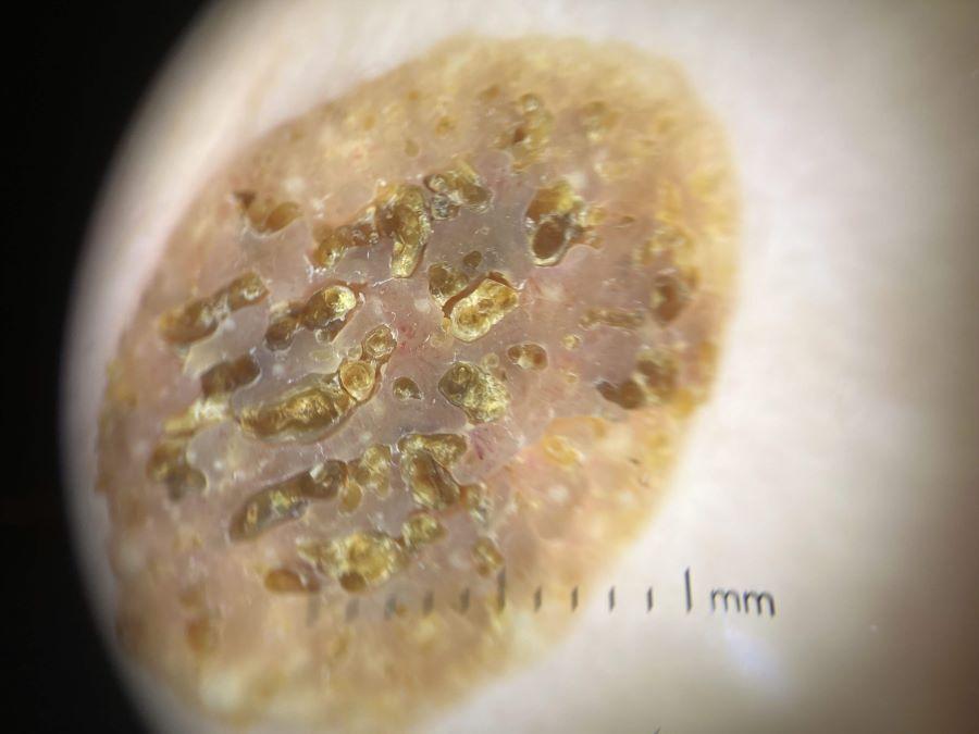
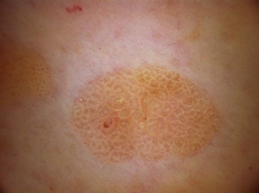
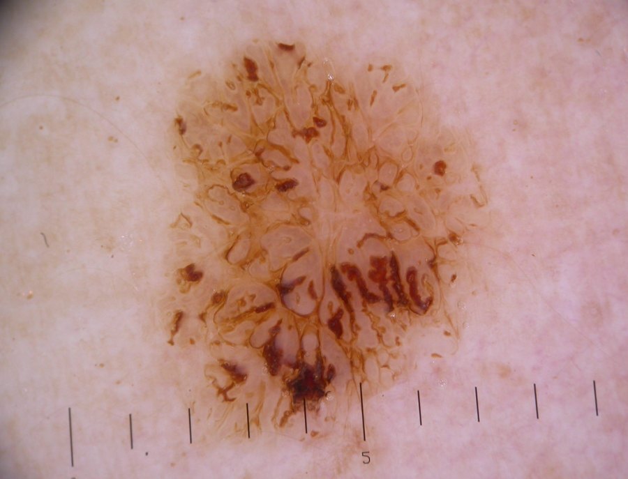
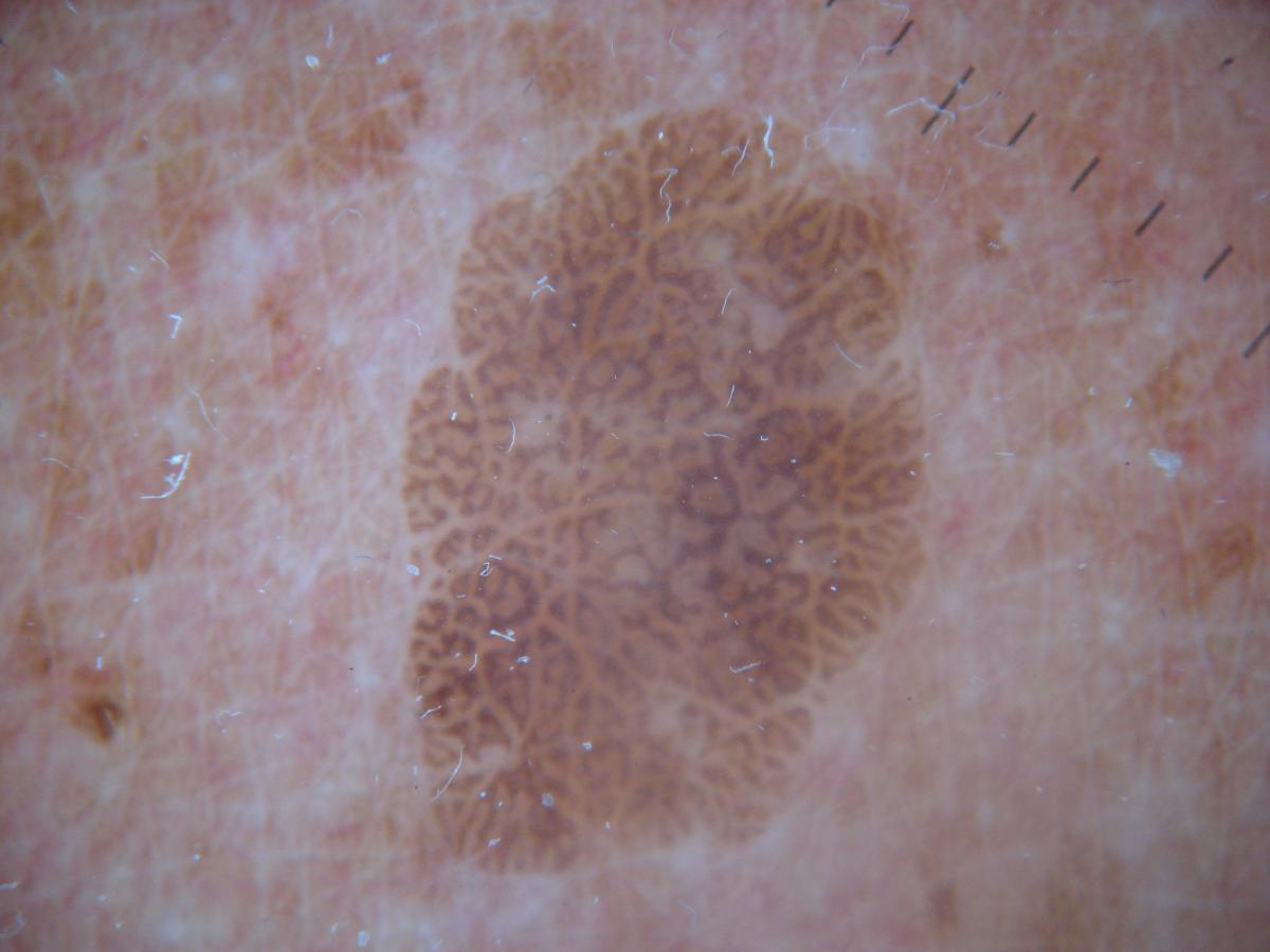
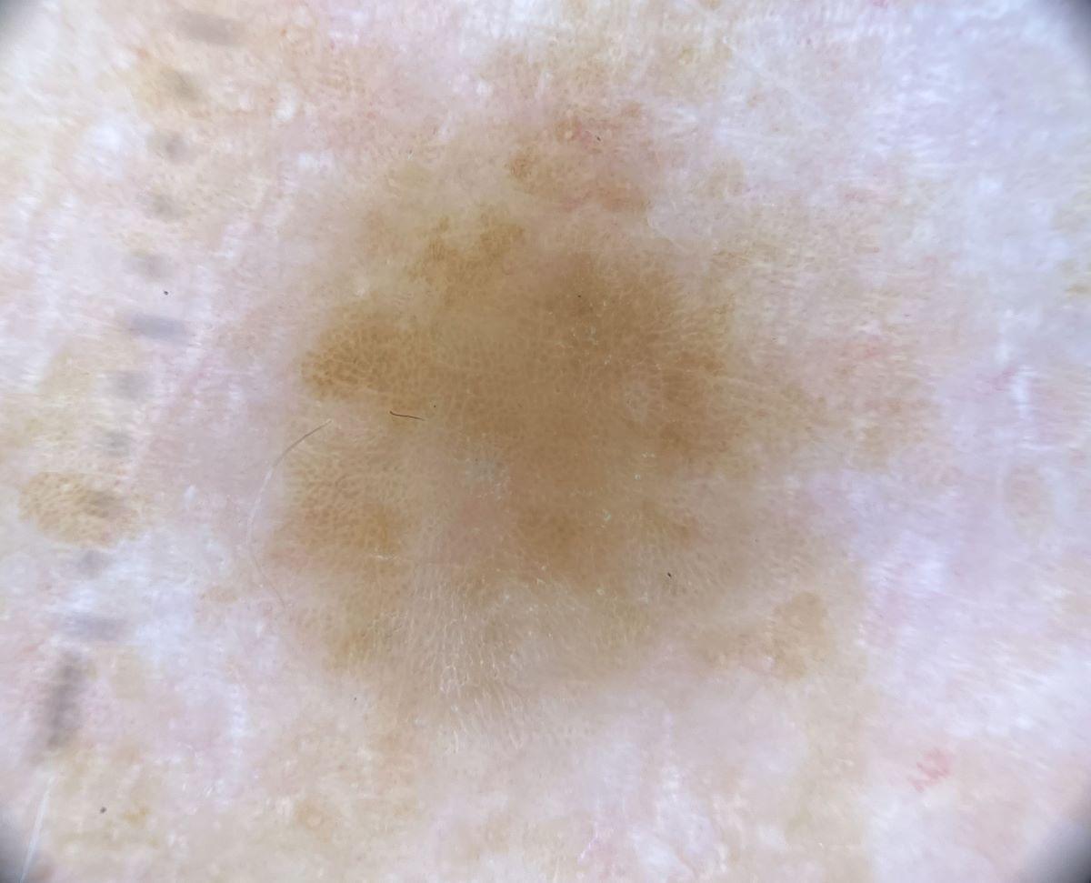
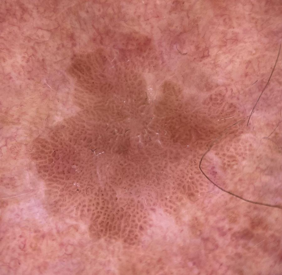
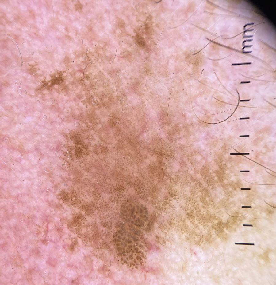
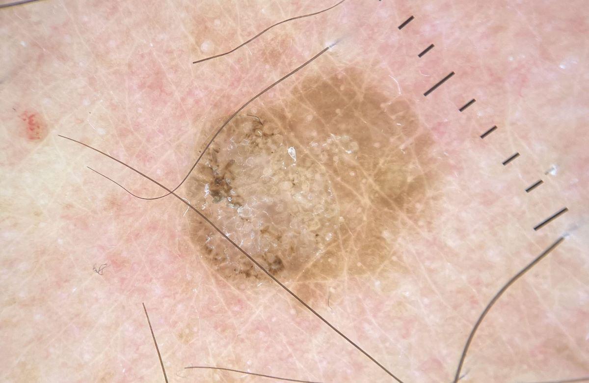
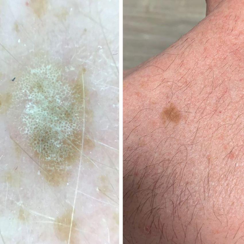
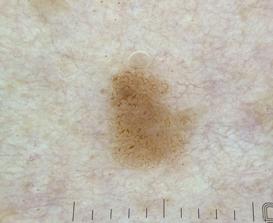
Seborrhoeic keratoses take on various appearances. The four most common patterns are:
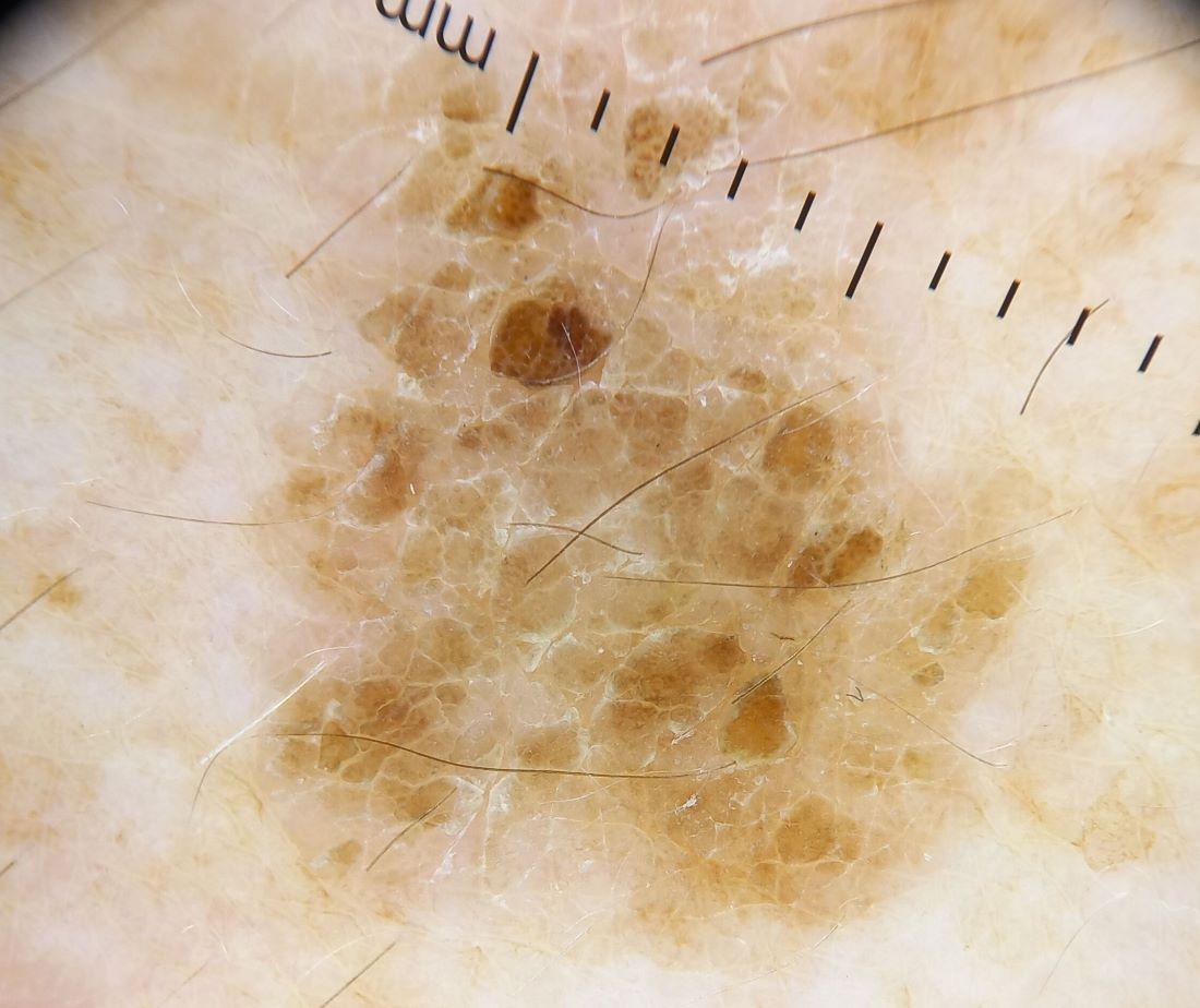
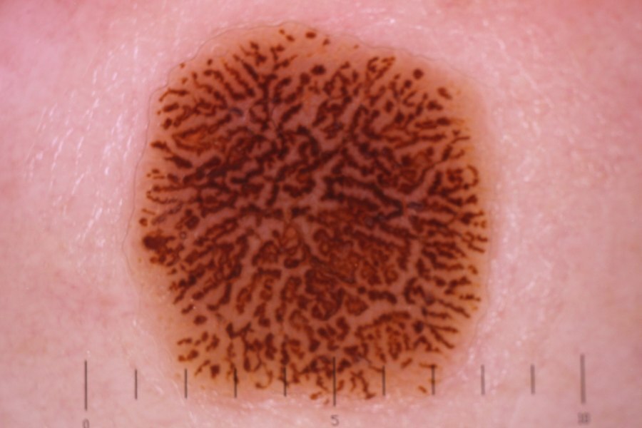
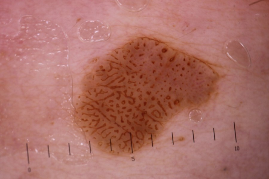
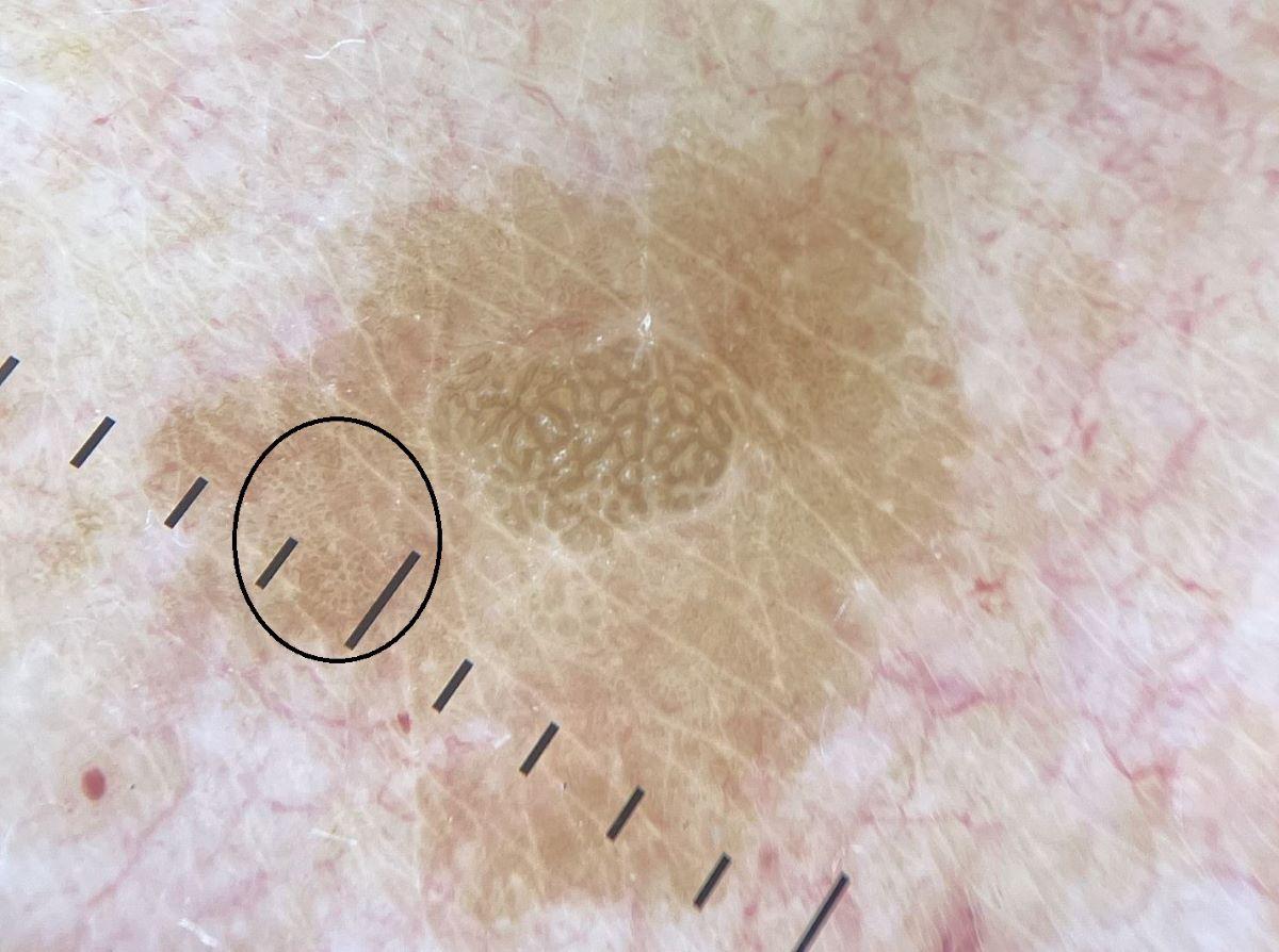
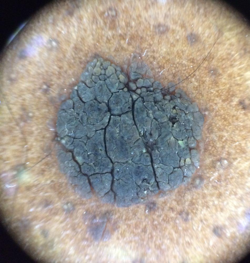
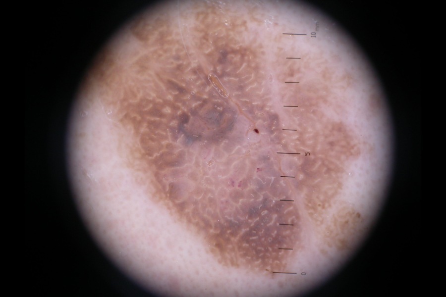
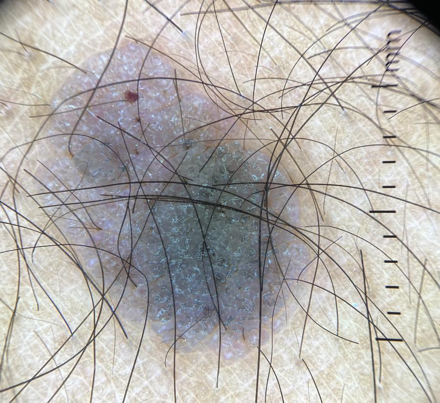
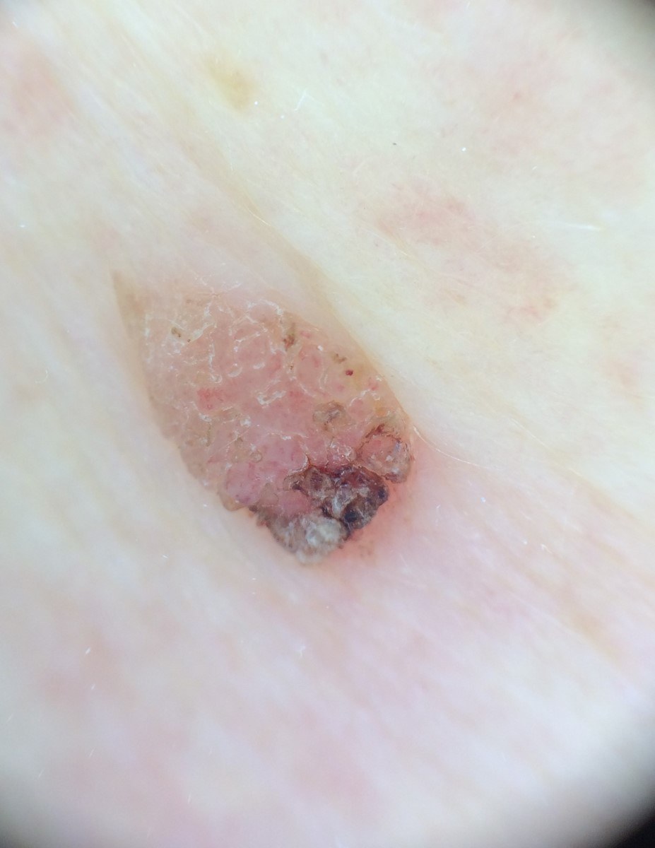
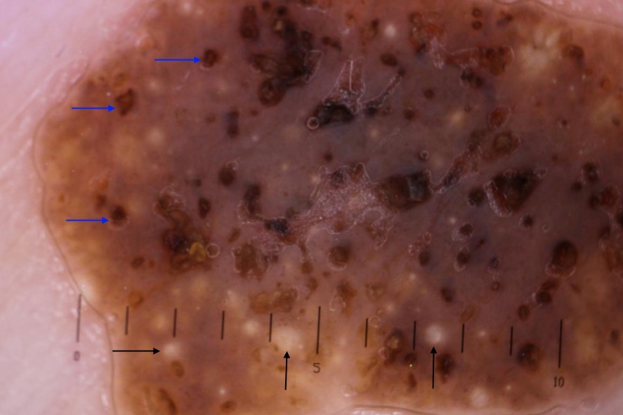
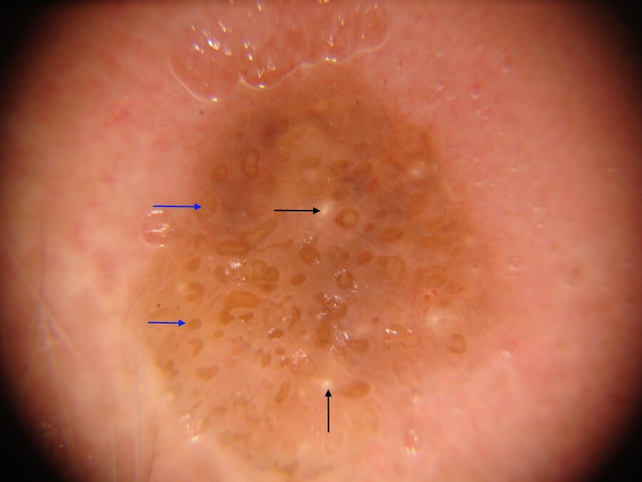
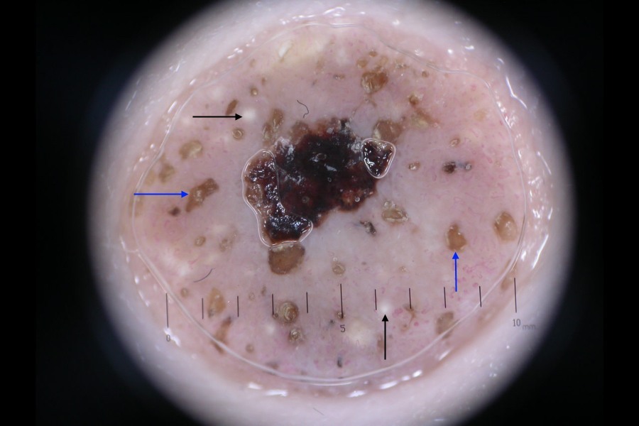
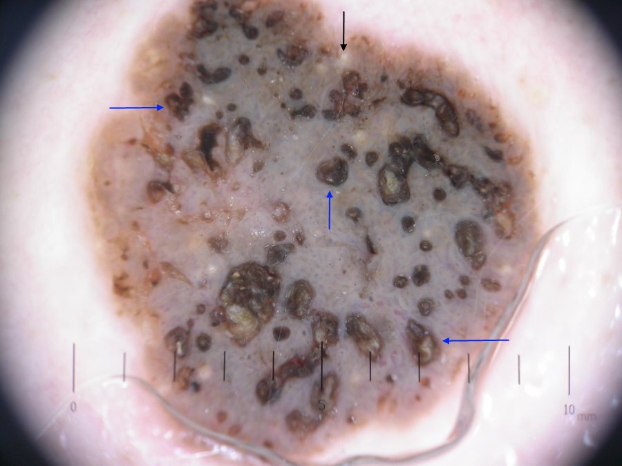
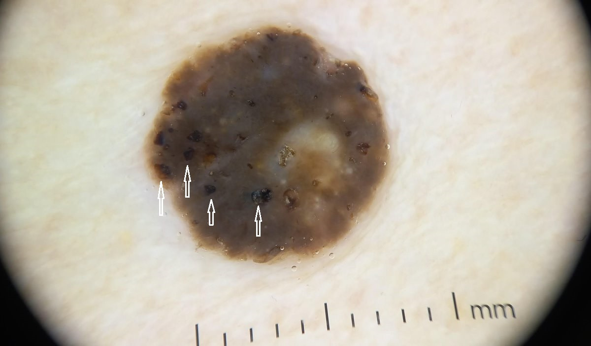
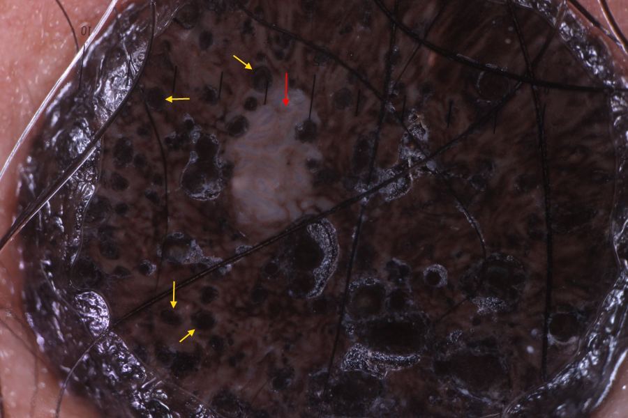
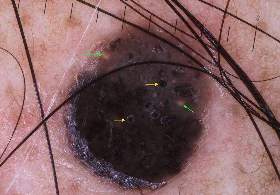
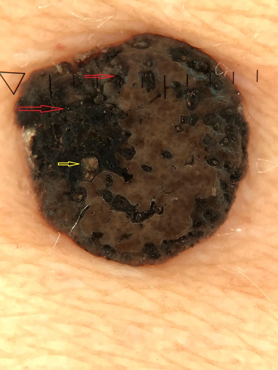
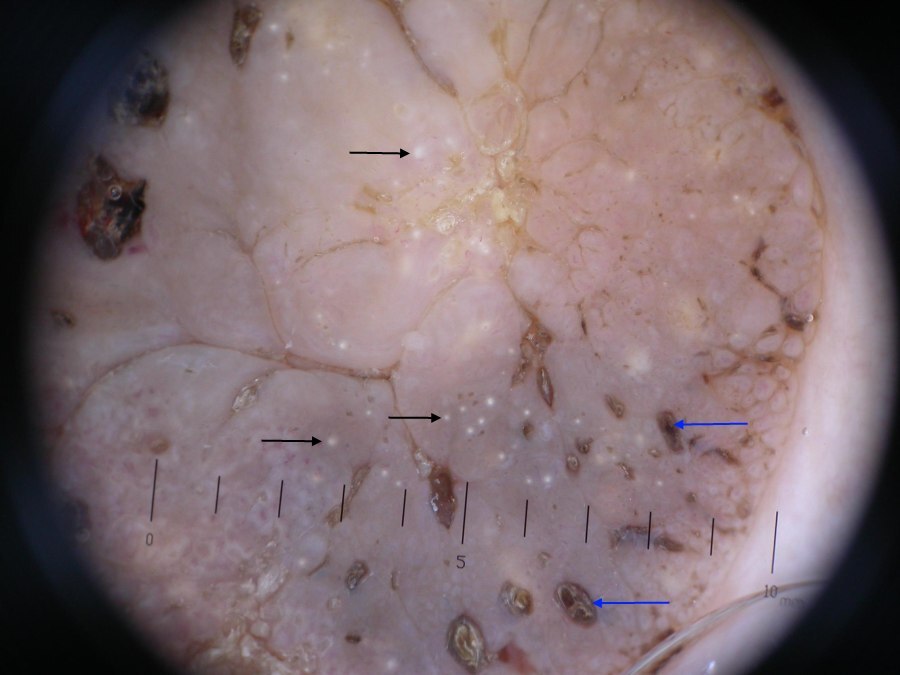
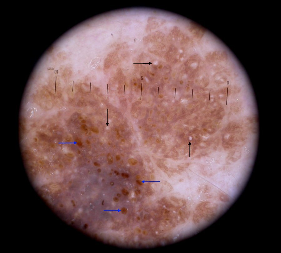
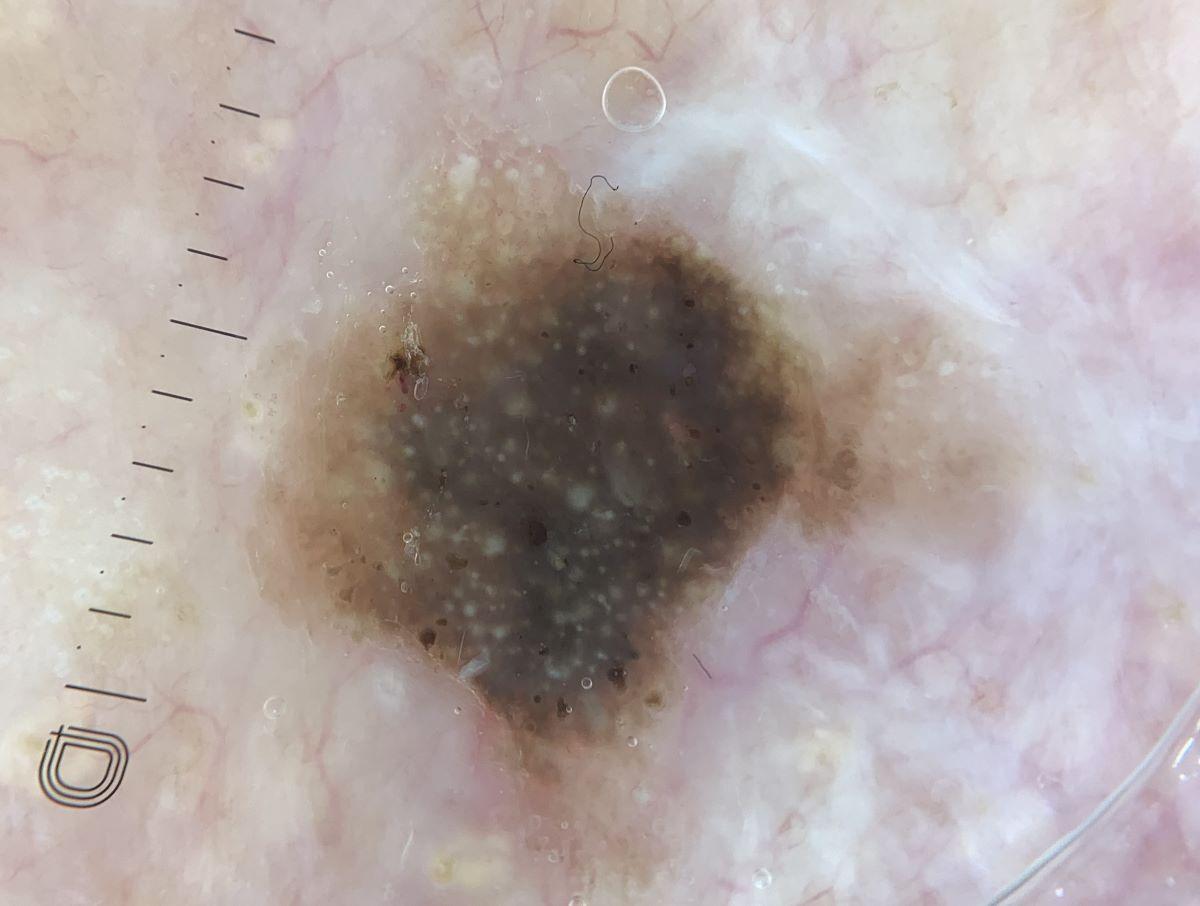
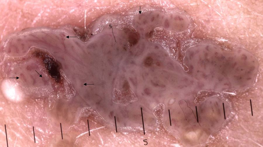
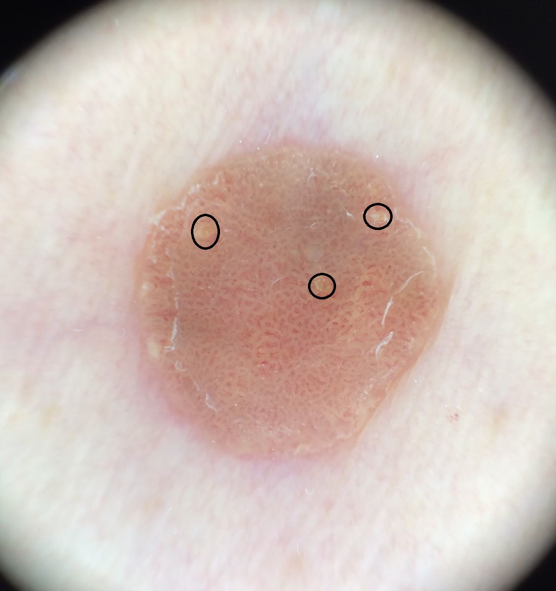
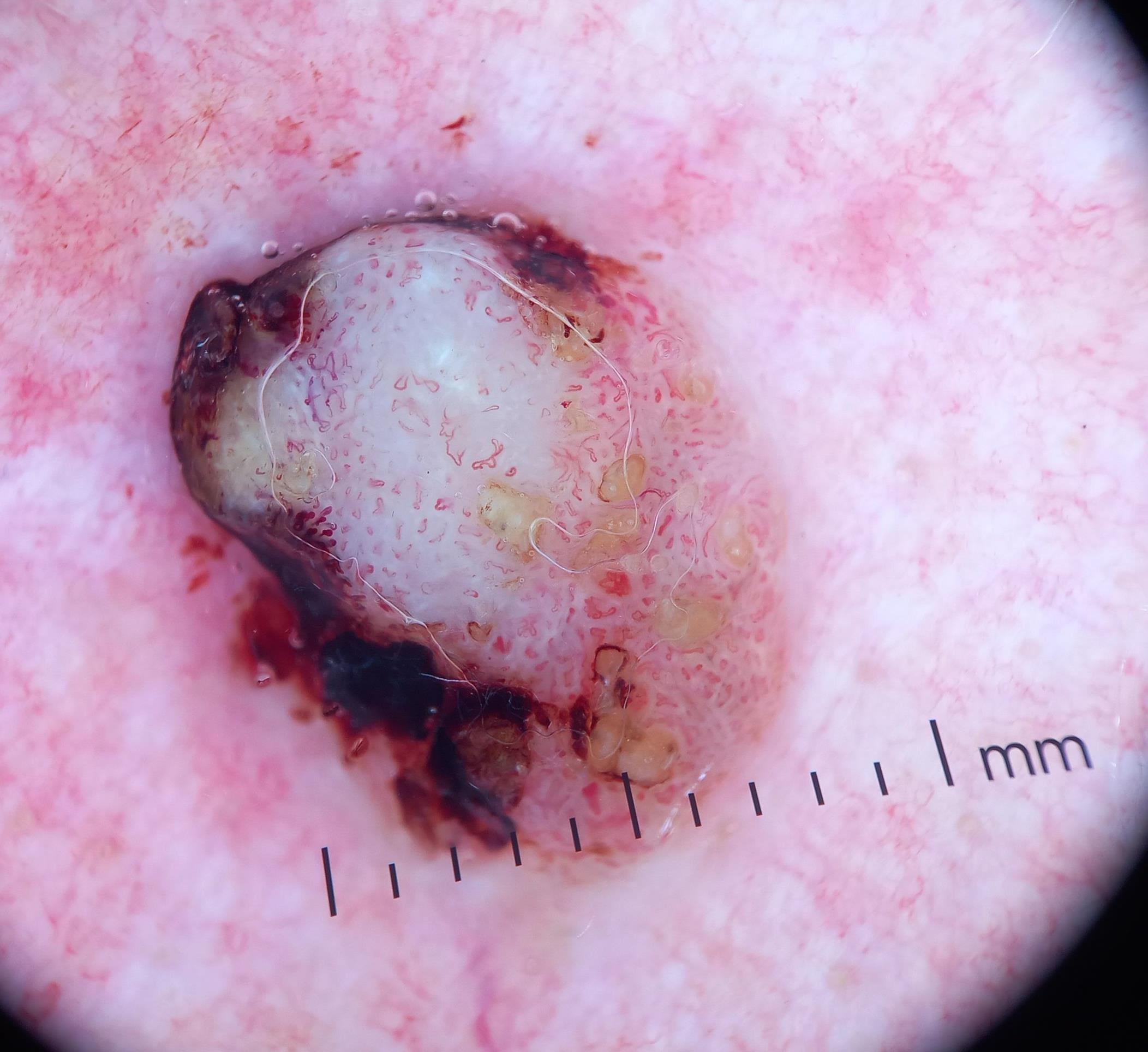
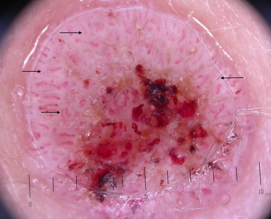
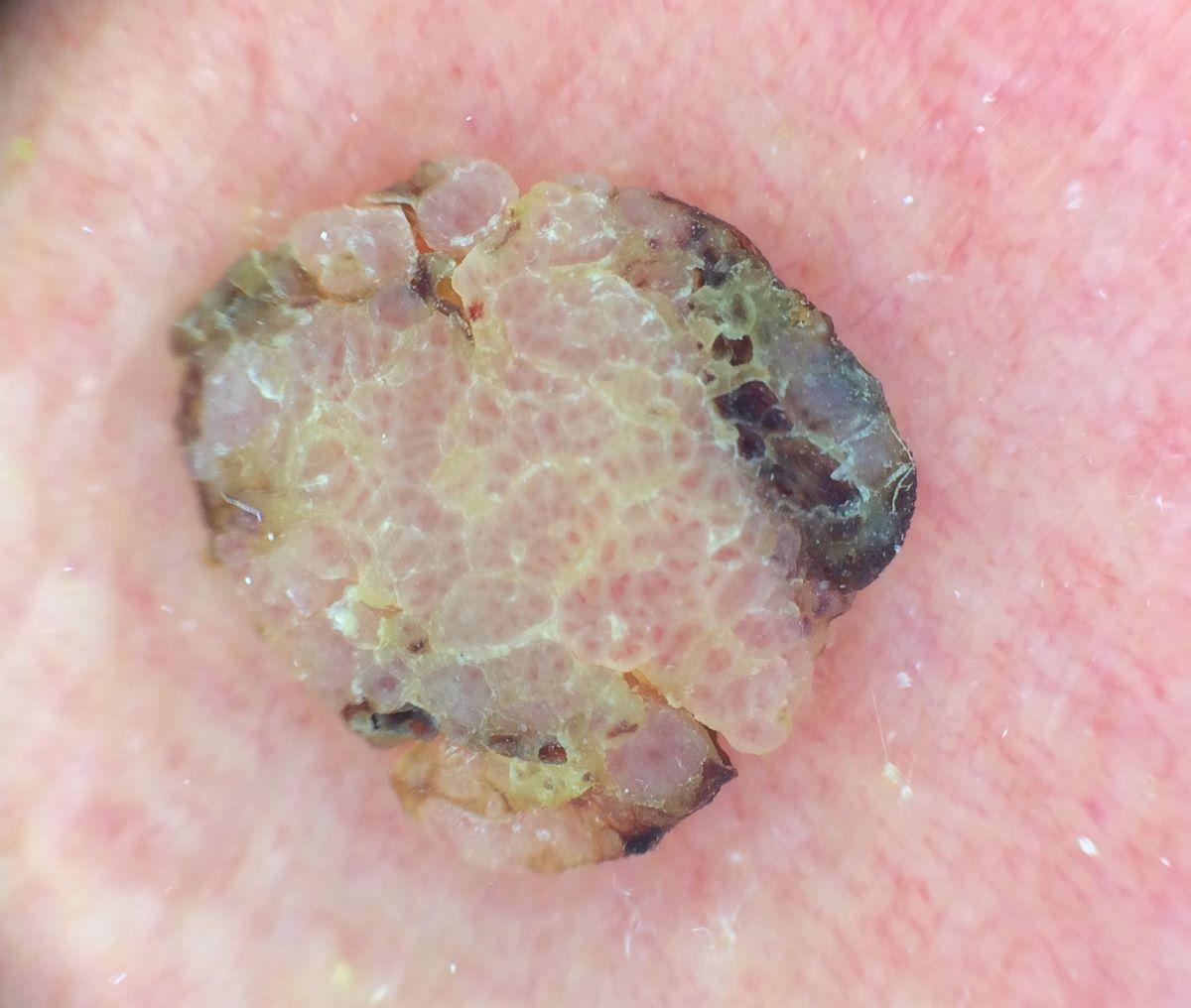
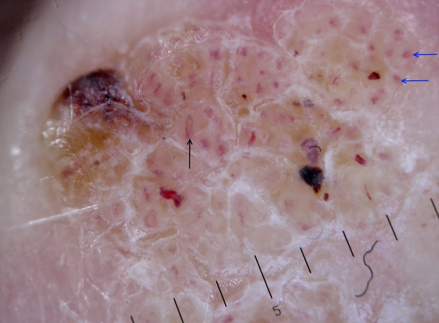
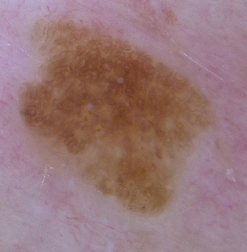
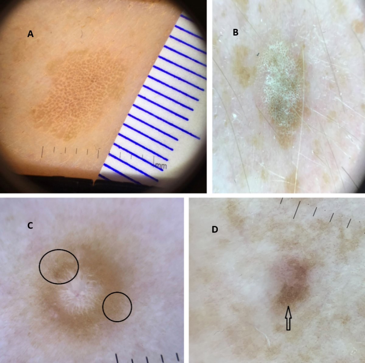
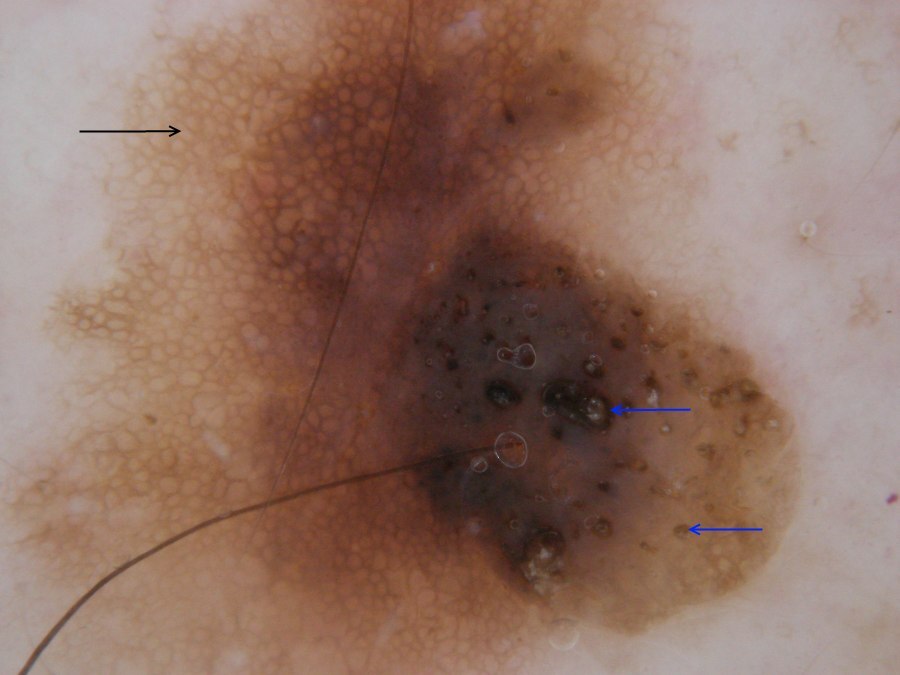
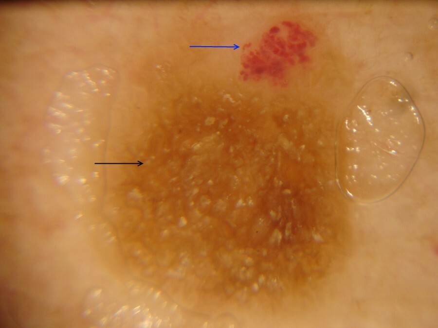
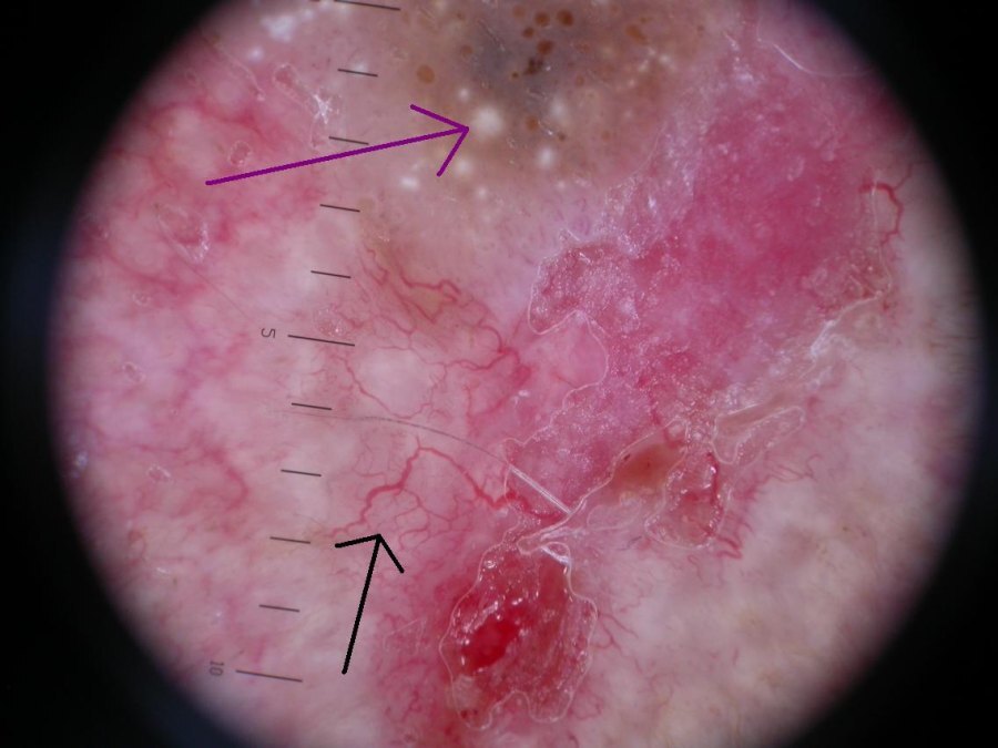
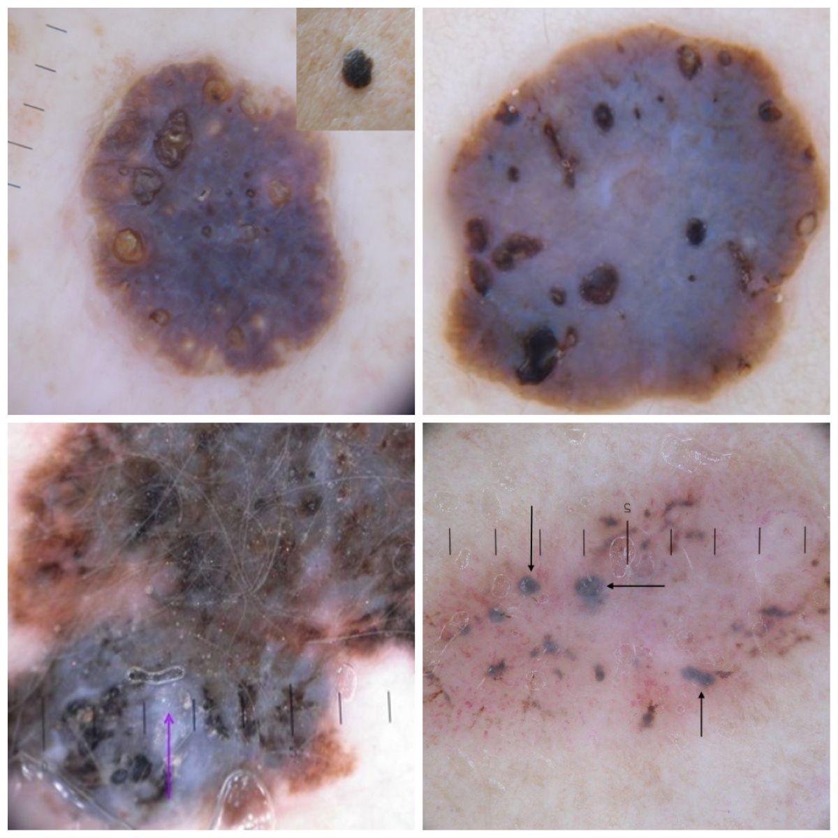
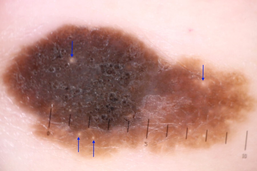
- Acanthotic lesions have a thickened epidermis with comedo-like openings (clods) and milia-like cysts. The comedo-like openings tend to be less regular in terms of shape and distribution compared to globules seen in melanocytic lesions, they can also appear more grainy as opposed to smooth. While milia-like cysts can appear in other conditions, multiple cysts (more than three) strongly suggest a benign non-melanocytic lesion, most commonly a seborrheic keratosis. Bloods vessels should be relatively uniform looped structures (for more information on vessels refer to the chapter Vessel patterns in dermoscopy)
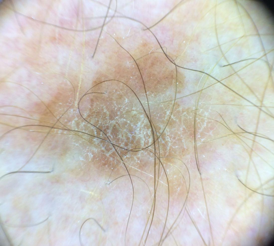
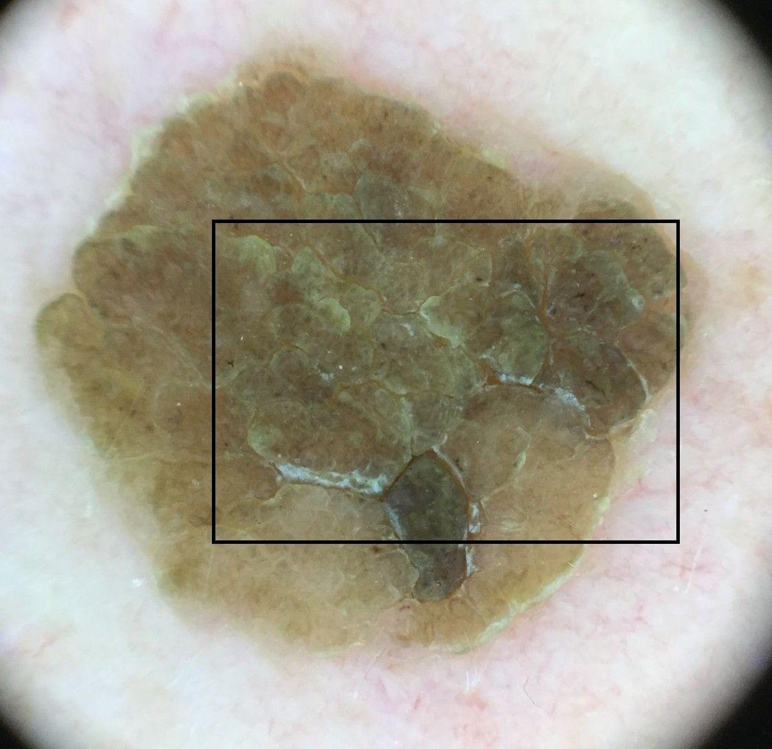
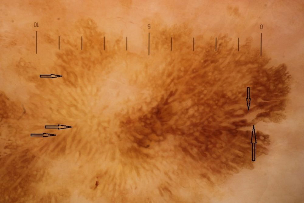
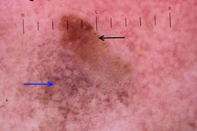
- Multiple fissures and ridges, sometimes with a cerebriform pattern (looking like the surface of the brain). Before lesions thicken the fissures and ridges have not yet developed and so appear as pigment bands (broader than lines, of variable length and width, are linear irregular or curved, and can be intertwined. Commonly yellow or brown). Alternatively, in very thin lesions some have small keratin scales that can build up in to a layered appearance
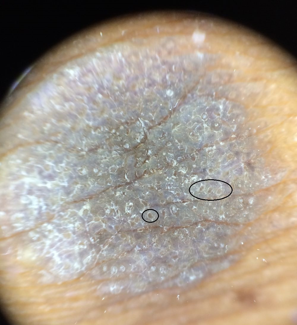
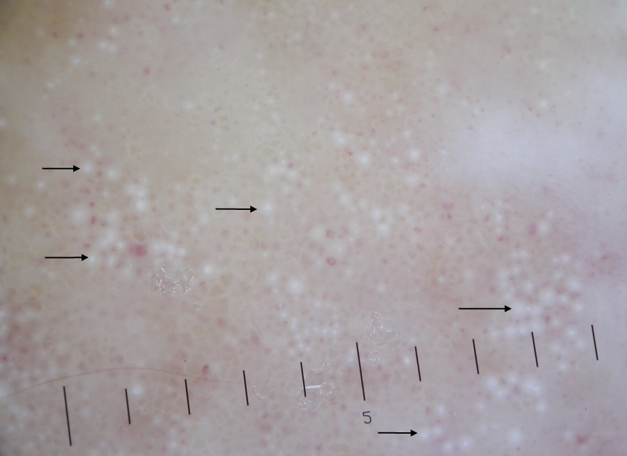
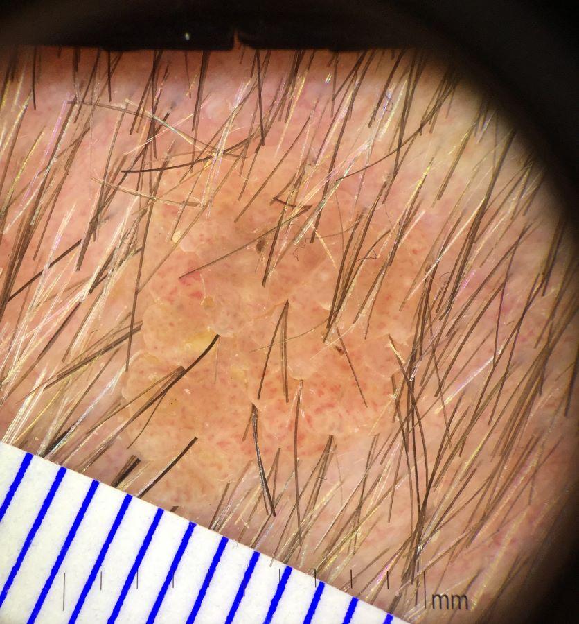
- Grouped skin-coloured clods with a central blood vessel - this pattern is mainly seen in pale lesions and gives a frogspawn-like appearance. Sometimes grouped clods are separated by fissures
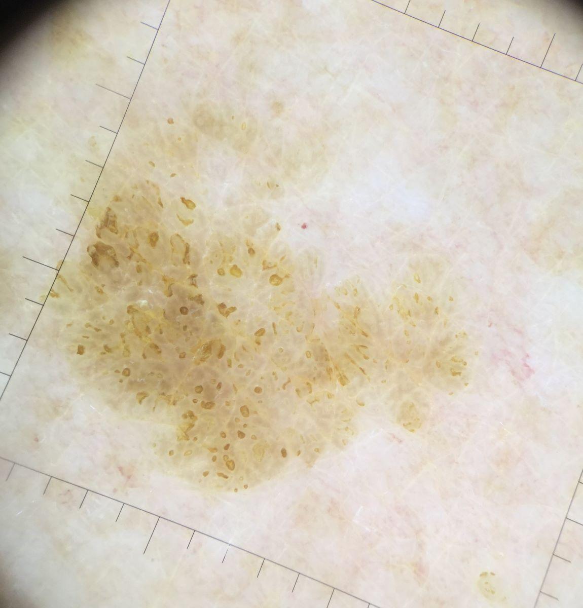
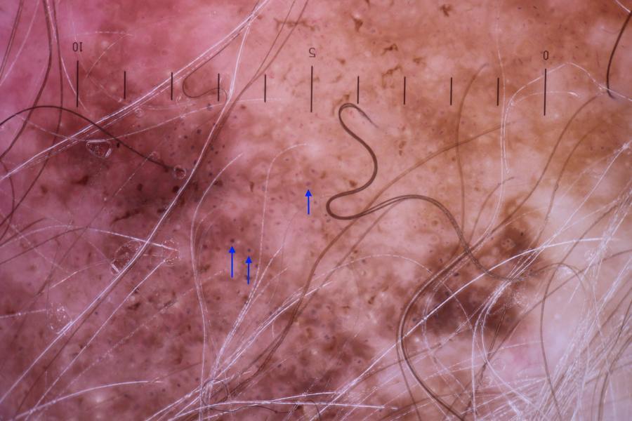
- Multiple small, brown, grouped, monomorphic (of the same or similar appearance) clods or circles - seen in some thinner lesions on the trunk and limbs. This is sometimes referred to as the 'clonal' pattern. Circles can also be found in dermatofibroma (at the periphery) and melanocytic lesions. The circles of seborrhoeic keratoses tend to be larger than those seen in melanocytic lesions
Dermoscopy is good for screening out many seborrhoeic keratoses; where doubt remains lesions should be referred for teledermoscopy or on an Urgent Skin Cancer Pathway.
Clinical Images
Please refer to notes on image rights at bottom of the page with regards to individual image
ownership.
1
2
3
4
5
6
7
8
9
10
11
12
13
14
15
16
17
18
19
20
21
22
23
24
25
26
27
28
29
30
31
32
33
34
35
36
37
38
39
40
41
42
43
44
45
46
47
48
49
50
51
52
53
54
55
56
57
58
59
60
61
62
63
64
65
66
67
68
69
70
71
72
73
74
75
76
77
78
79
80
81
82
83
84
85
86
87
88
89
90
91
92
93
94
95
96
97
98
99
Management
- Most seborrhoeic keratoses do not require treatment but for those that do the majority can be treated by liquid nitrogen (for more information refer to the chapter Cryosurgery). Very thick lesions are best removed by curettage and cautery - all samples should be sent for histology
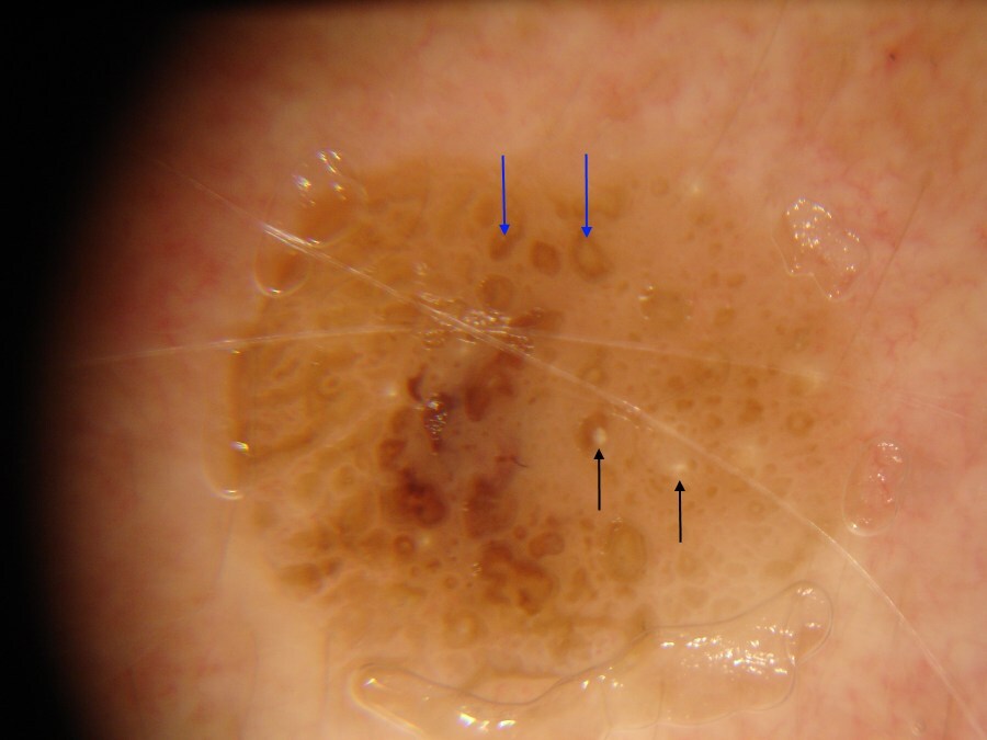
- If there is any uncertainty about the diagnosis the patient should be referred urgently for teledermoscopy or to Secondary Care (Urgent Skin Cancer pathway)
Disclaimer - the author PCDS cannot accept responsibility for any misleading or incorrect statements, and the management of individual patients remains the direct responsibility of the individual doctor. We do however hope that visitors to this site can contact us regarding comments that are considered misleading or incorrect so that we can continue to improve the site.
Image Rights - The PCDS would like to thank Dermatoweb, DermQuest (Galderma), and others who have contributed images. All named individuals and organisations maintain copyright for the relevant images.