Spitz naevi (including pigmented spindle cell naevus of Reed)

LAST UPDATED: Nov 17, 2025
Introduction

The Spitz naevus (syn. spindle cell naevus, epithelioid cell naevus, juvenile melanoma) is a variant of a melanocytic naevus, most commonly seen in children and young adultsMost authorities regard the pigmented spindle cell naevus of Reed (Reed naevus) a type of Spitz naevus. Although Spitz naevi are benign, they can have similar clinical and histopathological features to atypical Spitz naevi and melanoma (including Spitzoid melanoma), making their assessment difficult and best left in the hands of a specialist. 

This chapter is set out as follows:


History

  • Both lesions are very uncommon over the age of 30 years  

Clinical findings

History of Spitz naevi

  • Most common in children and young adults, 70% arise before the age of 20 years
  • After an early period of relatively rapid growth to reach an approximate size of 1 cm, lesions tend to become static around six months

Clinical features of Spitz naevi

  • Distribution
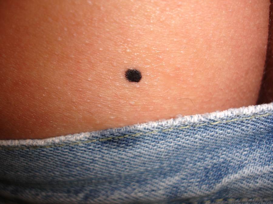
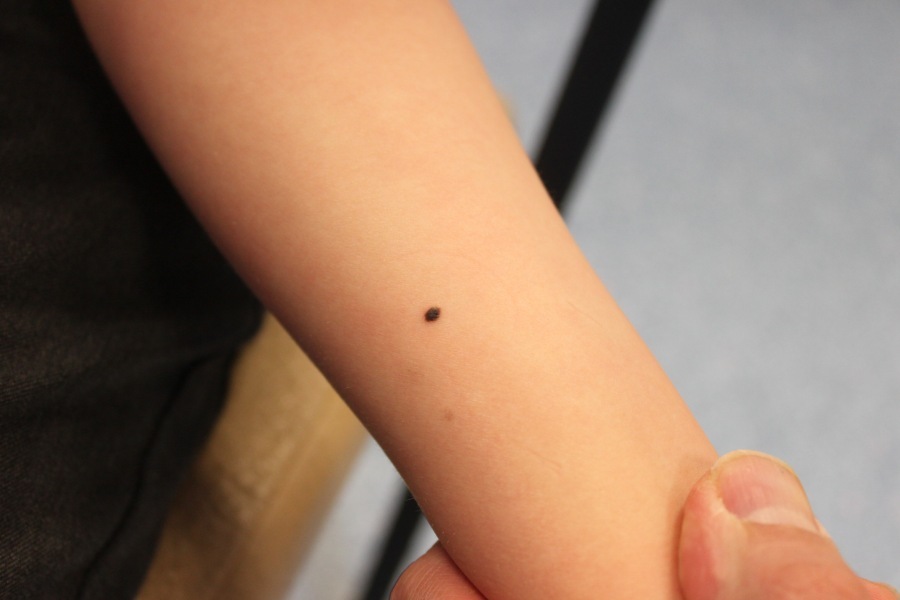
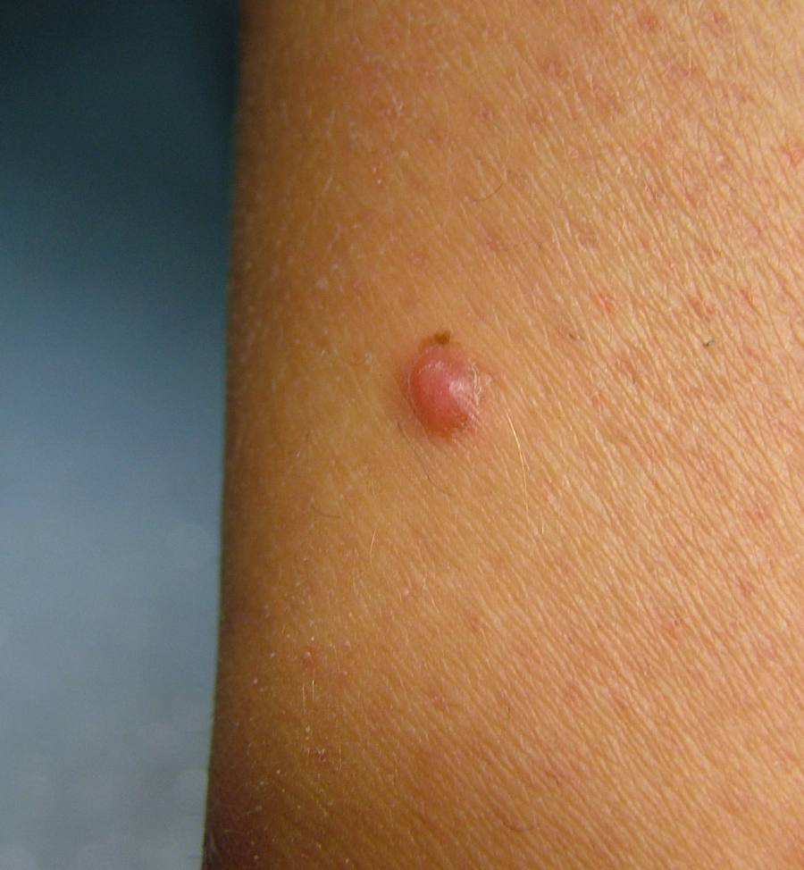
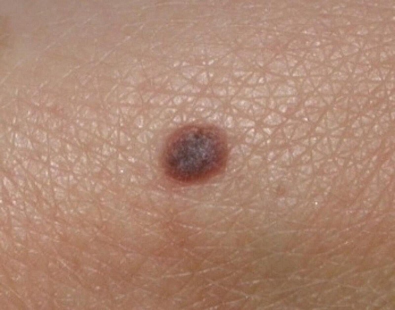
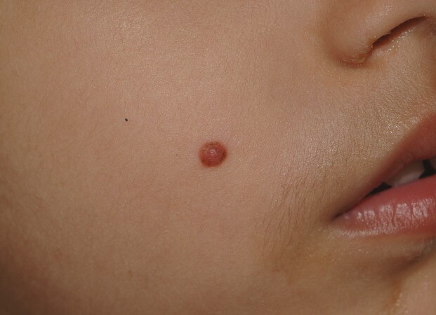
    • Any site. Classical Spitz naevi most commonly affect the face. Reed naevi most commonly affect the limbs
       
  • Morphology
    • Often an 'ugly duckling' ie different to the patient's other naevi
    • Usually solitary, multiple lesions very uncommon
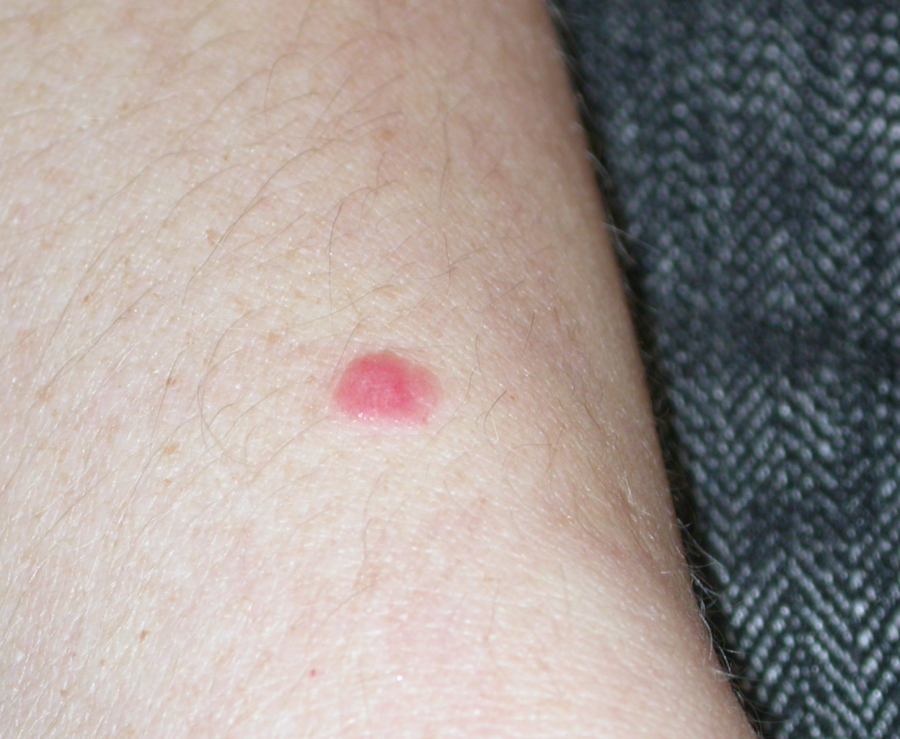
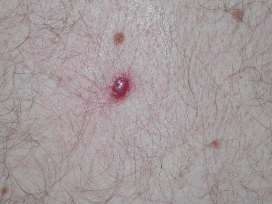
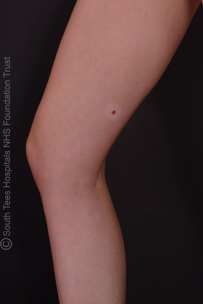
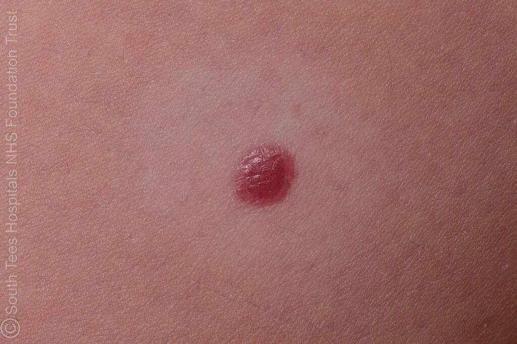
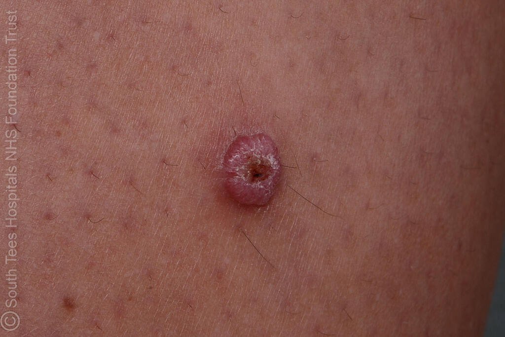
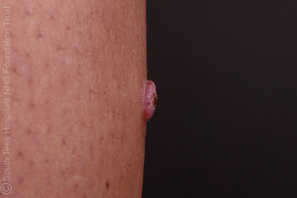
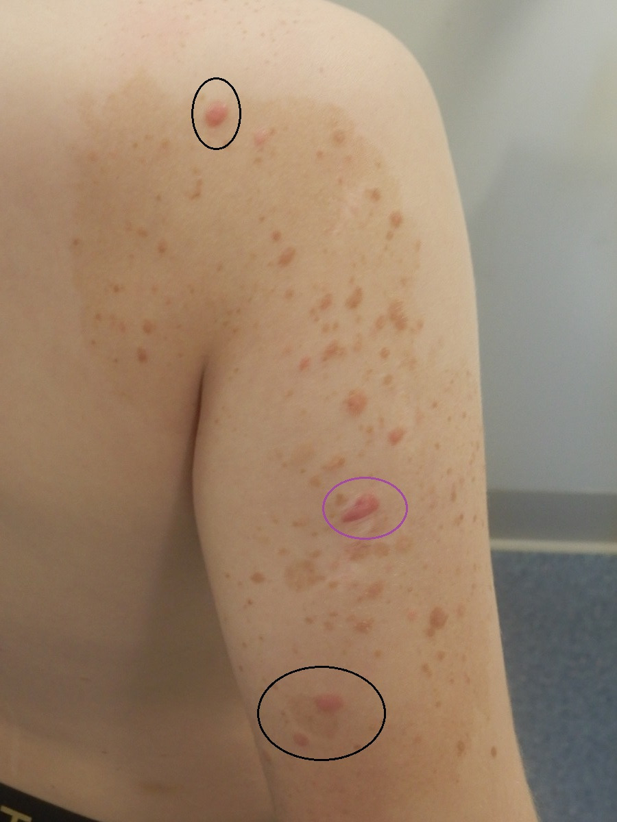
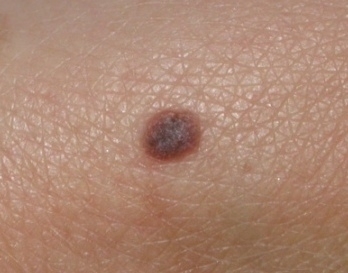
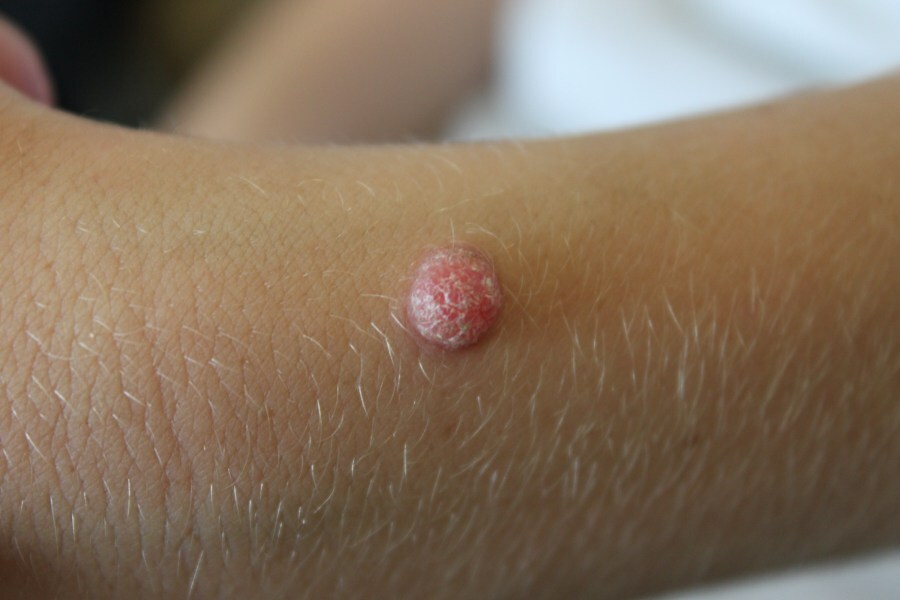
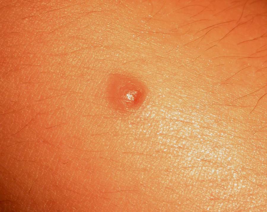
    • Classical Spitz naevi present as a raised firm papule or nodule, often pink-red but can be brown
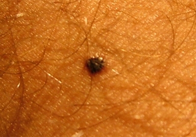
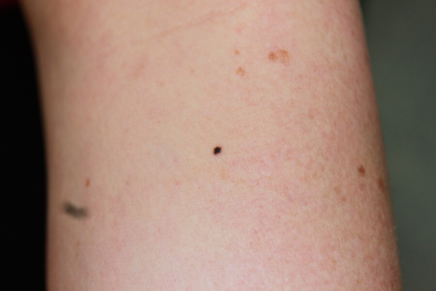
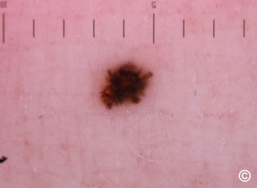
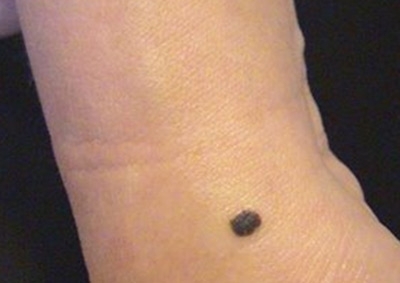
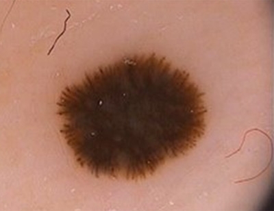
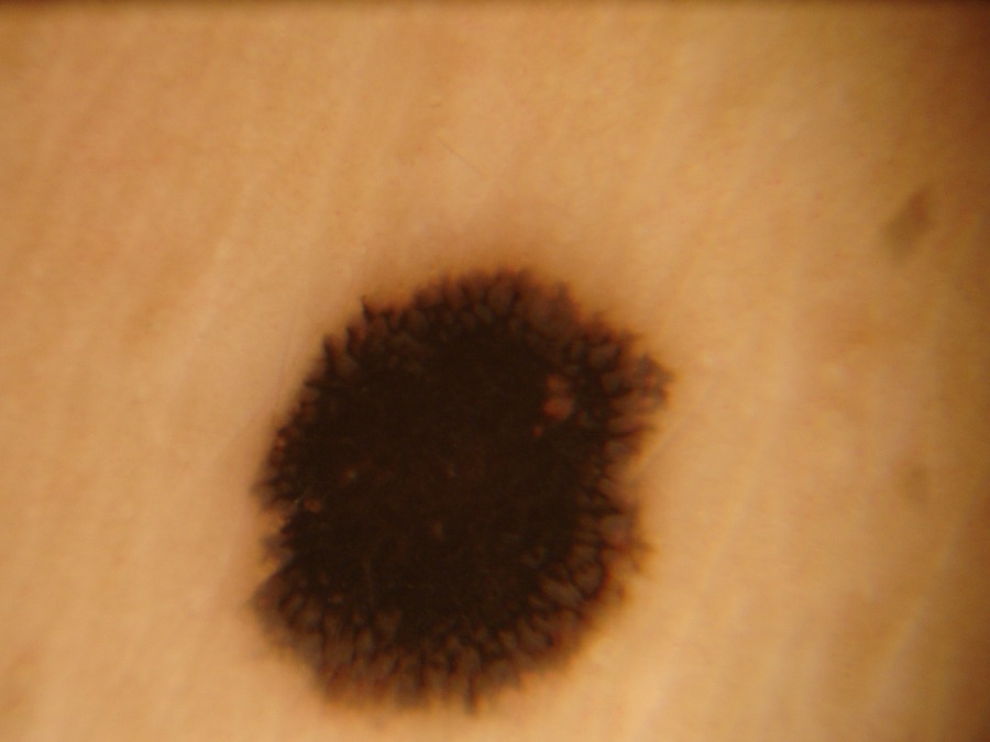
    • Reed naevi tend to be dark brown or black macules, or slightly elevated papules 

Dermoscopic features of Spitz naevi (hover over terminologies for description)

As with the clinical appearance, the dermoscopic appearance should be that of an 'ugly duckling' ie different to the patient's other naevi. Multiple patterns have been identified:

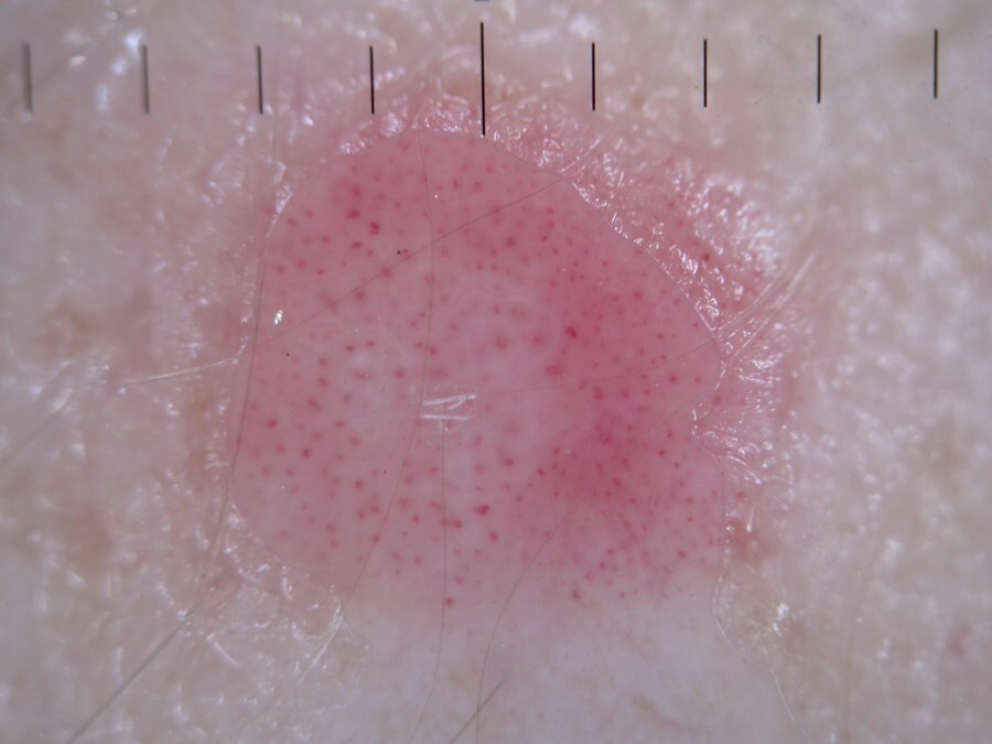
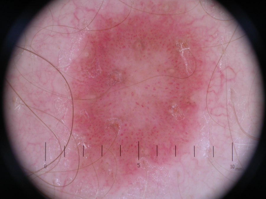
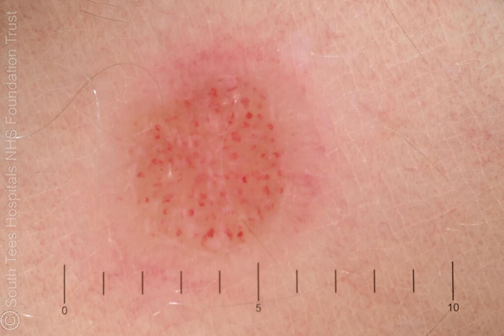
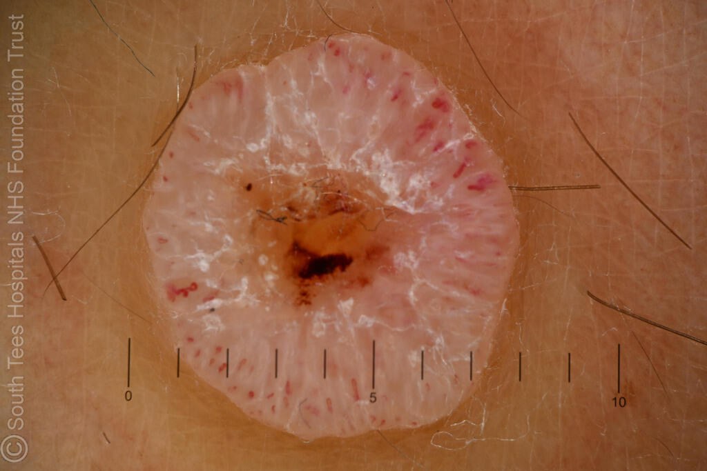
  • A regular dotted vascular pattern (click on the link for an overview of dermoscopic vessel patterns) is the hallmark of non-pigmented Spitz naevi. Another feature of non-pigmented Spitz naevi is the presence of shiny white structures (most commonly a white network or short white lines), best seen with polarised dermoscopy
     
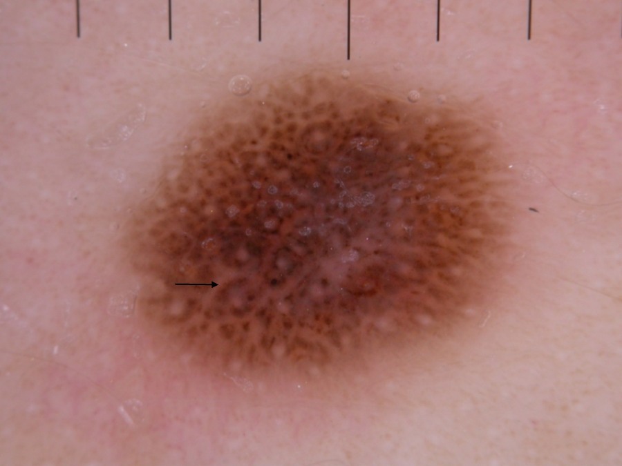
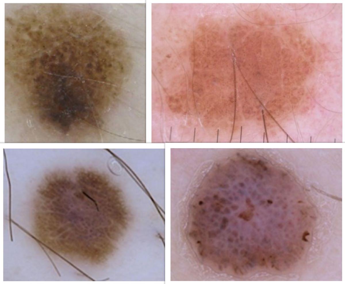
  • A pattern of globules evenly distributed throughout the lesion, sometimes with a relatively diffuse / symmetrical negative network (also known as a negative pigment network)
     
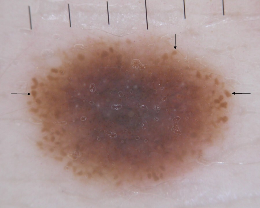
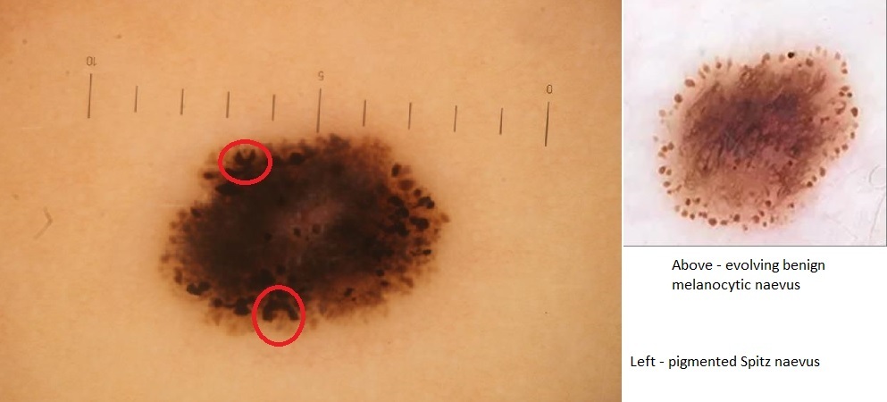
  • A largely structureless centre with layered (tiered) peripheral globules leading to a relatively symmetrical appearance. Some globules may appear as 'c-shaped'. In contrast, common benign melanocytic naevi tend to have a single layer of peripheral globules
     
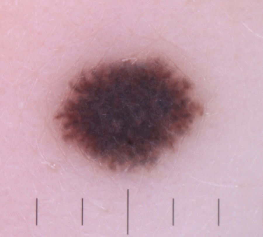
  • As with other Spitz naevi, the appearance of Reed naevi varies depending on their state of evolution:
    • Initially dark brown to black globules
    • The characteristic starburst pattern appears as the lesion evolves with streaks (radial lines) or pseudopods that are distributed symmetrically around the entire circumference
    • The centre becomes dark and structureless
    • Eventually, a progressive decrease of pigmentation is noted, or the lesion may completely involute

Features more suggestive of atypical Spitz naevi or melanoma

Suspected Spitz naevi should always be referred urgently to Secondary Care (2ww/USC pathway). Lesions are usually excised; histology forms should provide the pathologist with adequate clinical information and a clear differential diagnosis. Features more suggestive of an atypical Spitz naevus or melanoma include: 

  • Age - middle-aged and older patients
  • Colour - papules and nodules that are both pink and brown (sometimes the brown can be subtle)
  • Vascular pattern - unevenly distributed or polymorphous (ie more than one vessel type)
  • Globules - irregularly distributed, either throughout the lesion or at the periphery where the layers may be thicker in some areas. Increased variation in size, shape, colour
  • Peripheral streaks - asymmetrically arranged
  • Shiny white structures - increased asymmetry of lines including more in the way of orthogonal lines (perpendicular)
  • Negative network - focal and/or asymmetrical distribution 

Clinical Images

Please refer to notes on image rights at bottom of the page with regards to individual image ownership.
Investigations

Histology

  • In Spitz naevi cells may be spindle-shaped or epithelioid and arranged in clusters. Kamino bodies are found at the dermo-epidermal junction, but these are not specific for a Spitz naevus as they can also be found in early melanoma. In Reed's naevi large quantities of melanin are found in the naevus cells
     
  • The following histopathological features may be found in a Spitzoid melanoma:
    • Upwards migration of naevus cells into the epidermis (pagetoid spread)
    • Pagetoid spread at the edge or beyond the lateral margin of the naevus
    • Although there may be a few mitotic figures in a Spitz naevus, the presence of larger numbers of mitotic figures is suspicious
    • Any mitotic figures in deeper cells
    • Any abnormal mitotic figures
       
  • Follow these links for more detailed histological features of atypical Spitz naevi and Spitzoid melanoma

Management

  • Spitz naevi can mimic melanoma, clinically, dermoscopically and histopathologically, and should be referred urgently to Secondary Care (2ww/USC pathway) 
     
  • In Secondary Care lesions are generally managed by excision with a 2 mm margin of normal surrounding skin. Occasionally, some specialists observe flat Reed naevi in patient's aged under 12 years. All raised Spitz or Reed naevi are excised regardless of age, since clinically one cannot differentiate a typical from an atypical Spitz nor Spitzoid melanoma at any age
     
  • Atypical Spitz naevi (sometimes referred to as Spitz tumours of unknown malignant potential, or 'STUMP') are managed as per MDT decision (individualised as per dermpath and genetic markers), and Spitzoid melanoma as melanoma

Disclaimer - the author PCDS cannot accept responsibility for any misleading or incorrect statements, and the management of individual patients remains the direct responsibility of the individual doctor. We do however hope that visitors to this site can contact us regarding comments that are considered misleading or incorrect so that we can continue to improve the site.

Image Rights - The PCDS would like to thank Dermatoweb, DermQuest (Galderma), and others who have contributed images. All named individuals and organisations maintain copyright for the relevant images.

Quick Links

The following pharmaceutical companies have had no involvement in the content of this website or in our conference programmes


Almirall
Galderma
Glenmark
Johnson & Johnson
La Roche-Posay
LEO Pharma
Pierre Fabre
Schuco