Basal cell carcinoma: superficial

LAST UPDATED: Jan 19, 2026

Patient Information Leaflet
Link: Basal cell carcinoma

https://www.pcds.org.uk/patient-info-leaflets/basal-cell-carcinoma
Introduction

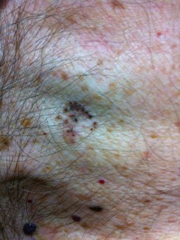
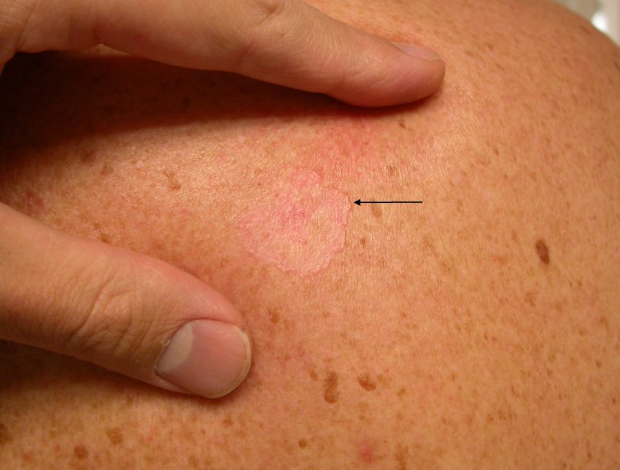
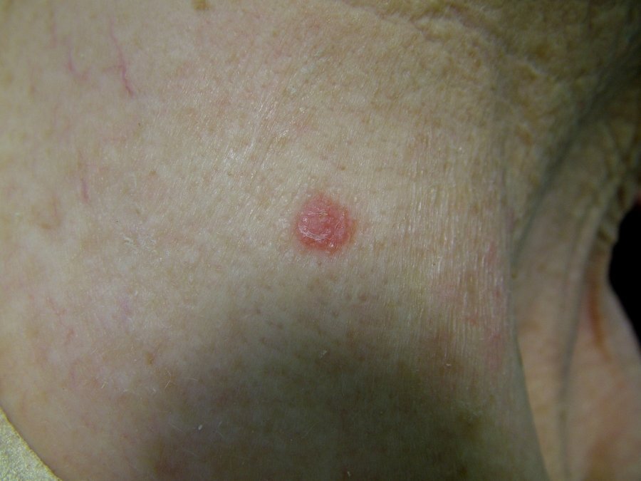
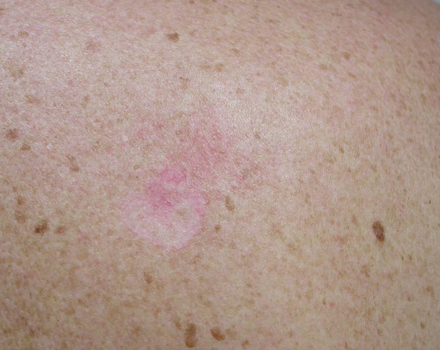
Superficial basal cell carcinoma (BCC) are the most common type of BCC in younger adults, and present most frequently on the trunk and shoulders as a slowly expanding pink-red patch. 

This chapter is set out as follows:


Clinical findings

Distribution

  • Frequently multiple
  • Most often found on the trunk, shoulders, and shins

Morphology

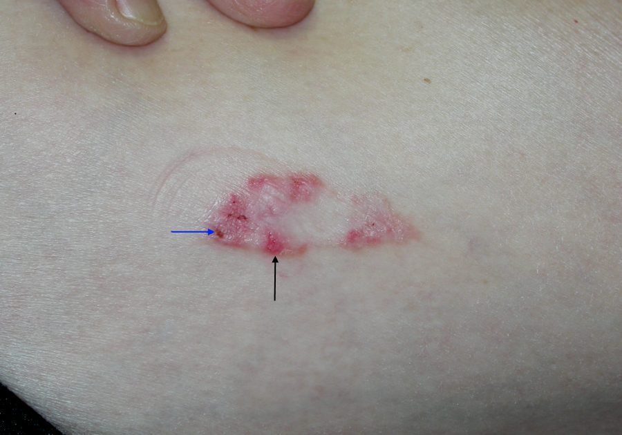
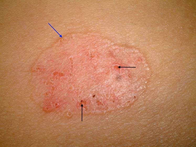
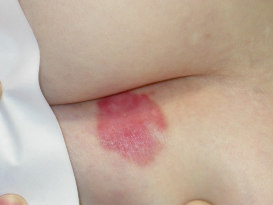
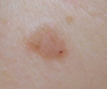
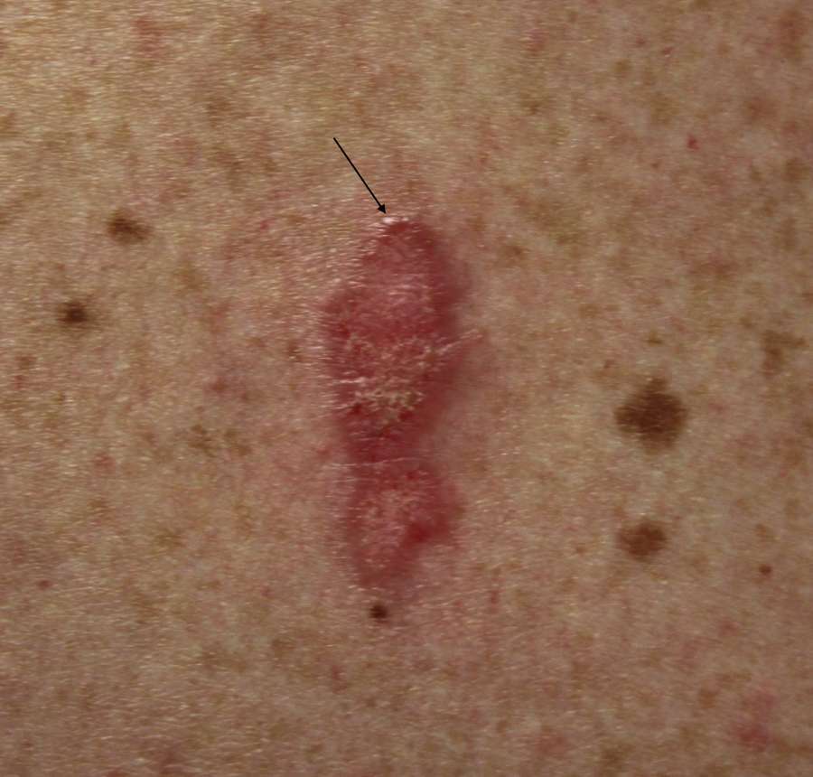
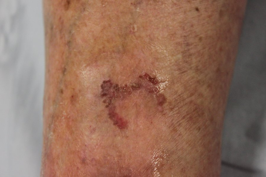
  • Expand slowly as a red / pink patch
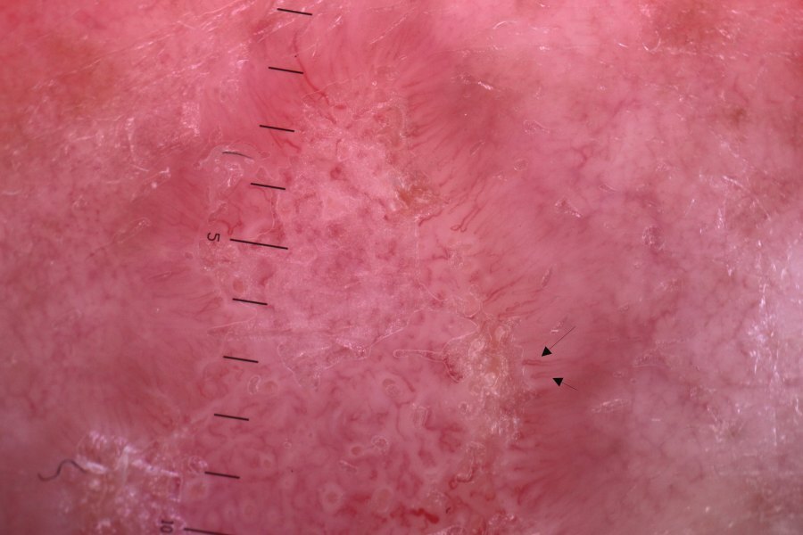
  • Develop a very fine raised 'whipcord' edge
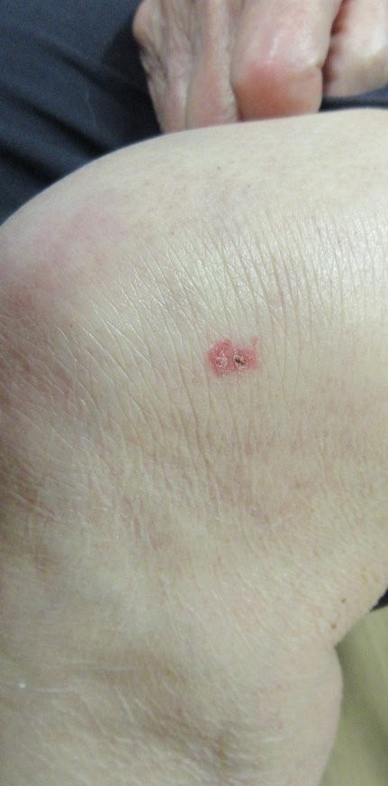
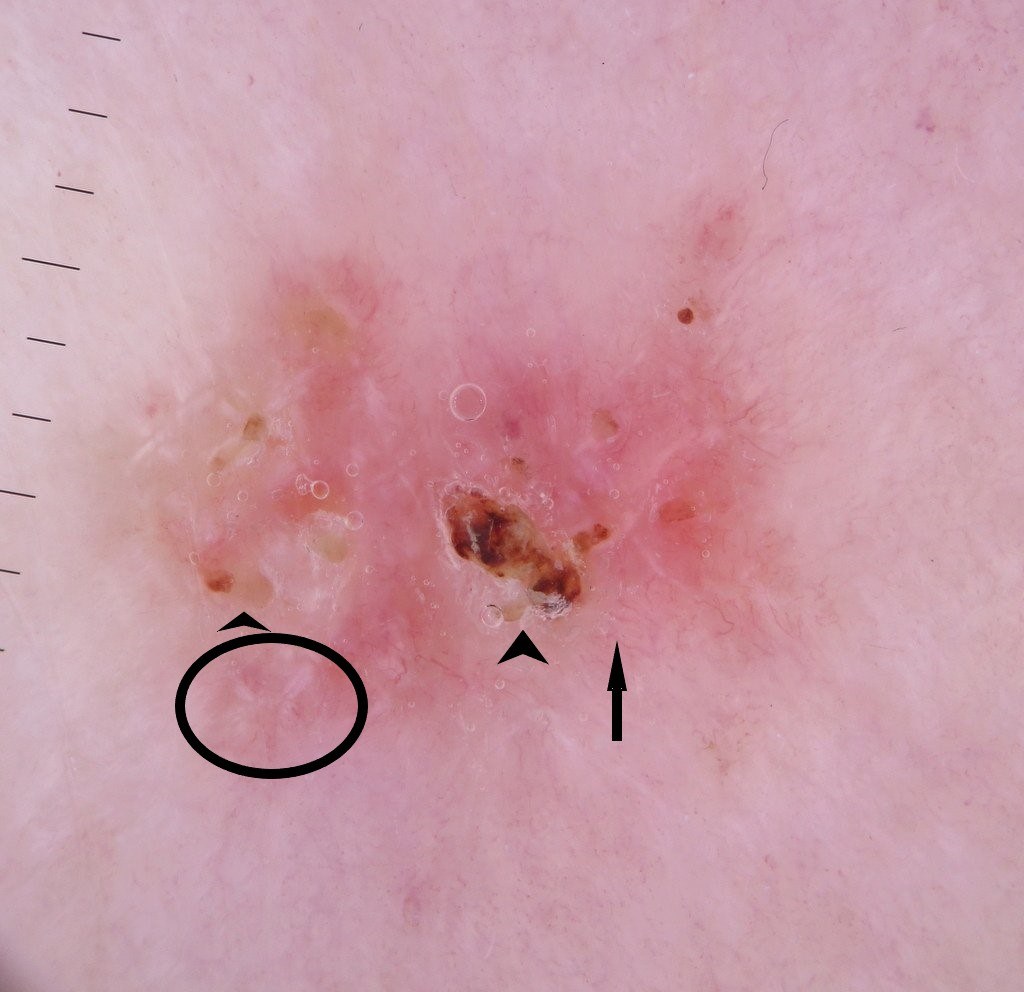
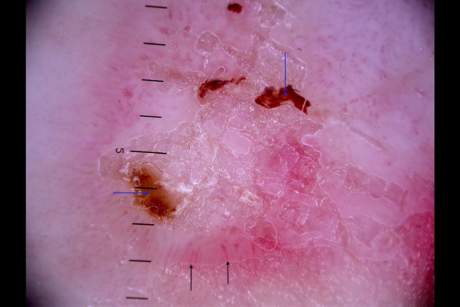
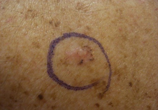
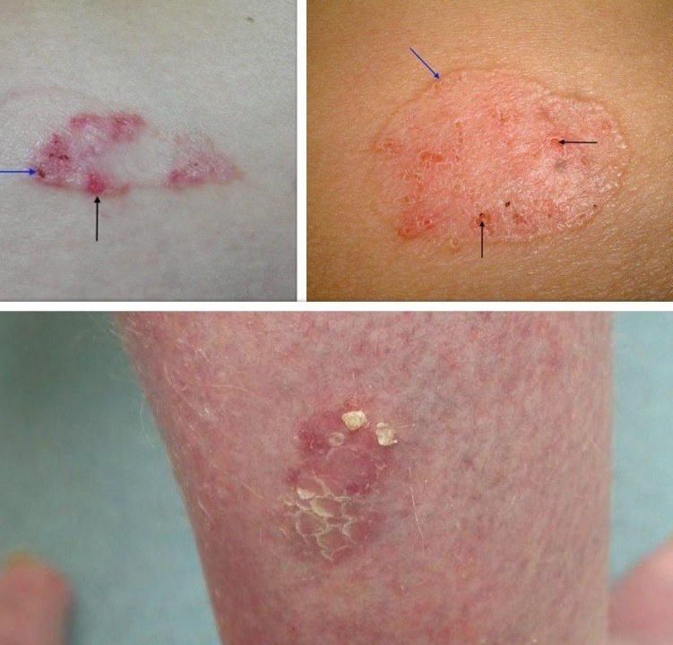
  • As they enlarge the surface becomes more fragile developing focal erosions and crust - a crust represents dried blood, plasma, or exudate that results from the erosion. Erosions / crust tend to have a different appearance to the white-yellow rough surface scale seen in actinic keratoses and Bowen's disease 

Dermoscopic features (hover over terminologies for description)

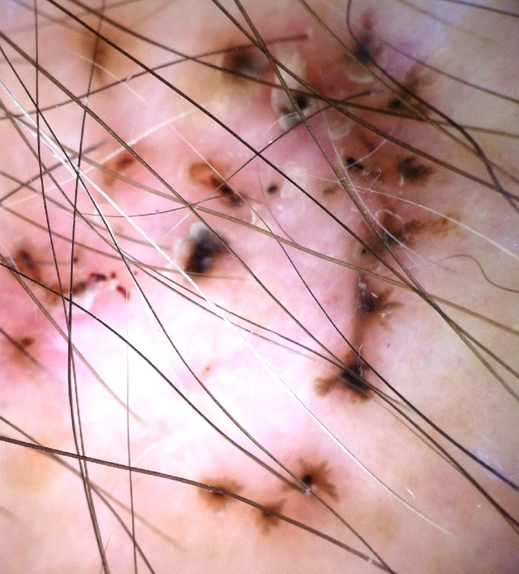
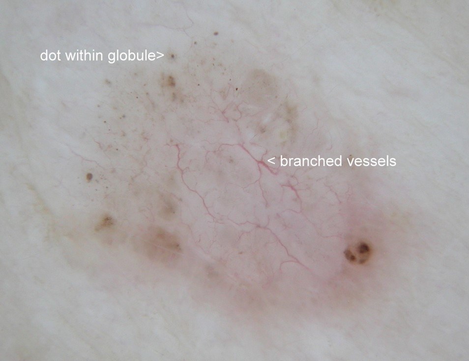
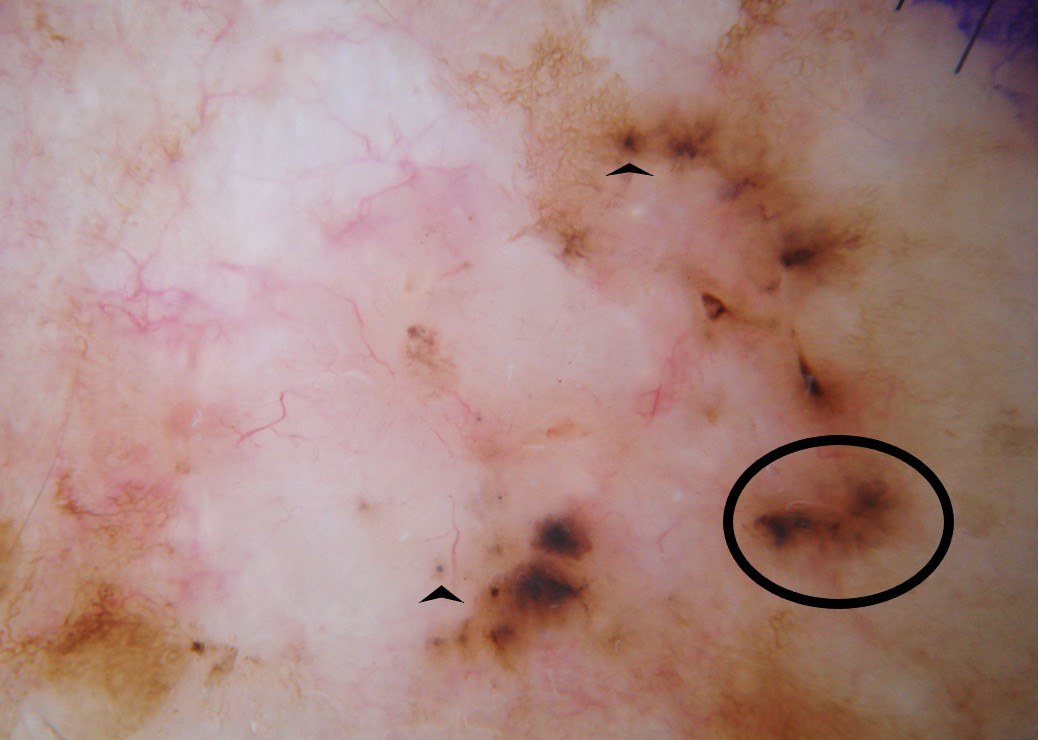
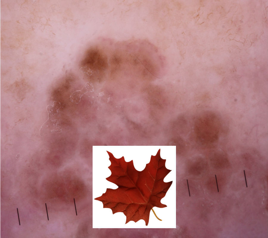
  • Focal erosions
     
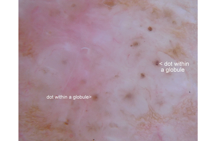
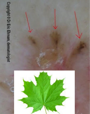
  • Much of the pigment in superficial BCC is peripheral:
  • Structureless white / red areas
     
  • Shiny-white structures
     
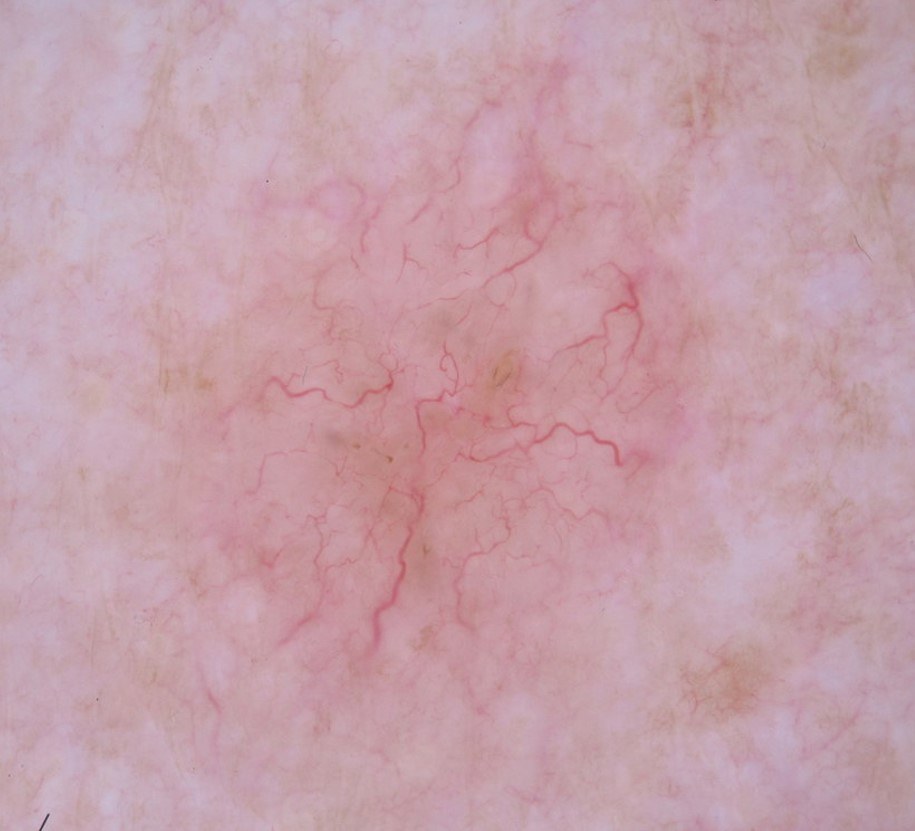
  • Vascular appearance - vessels are fine and sharply focused. They can be serpentine or short-fine branching vessels (narrower than typical arborising vessels of a solid BCC in which vessels tend to start off with a wider calibre that gets narrower as the vessels branch); occasionally peripheral parallel finely-looped vessels (for more information on vessels refer to the chapter Vessel patterns in dermoscopy)
     
  • Features against the lesion being a superficial BCC (favouring a solid BCC) - ulceration, arborising vessels, blue-grey clods

Clinical Images

Please refer to notes on image rights at bottom of the page with regards to individual image ownership.
Management

Patients need to be provided with options that include, in no particular order:

  • No treatment - may be the best choice for asymptomatic lesions on the trunk in patients with a reduced life expectancy
     
  • Topical treatments (to the lesion and 4 mm of normal surrounding skin)
    • 5% 5-fluouracil cream BD for 6 weeks
    • Imiquimod 5% cream (Aldara ®) 5 times a week for 6 weeks
       
  • Cryosurgery - only suitable for small lesions (two freeze-thaw cycles of 30 seconds to the lesion and 4 mm of normal surrounding skin)
     
  • Surgery - excision with a 4 mm margin or double curettage & cautery 
     
  • Photodynamic therapy

For  more information on the management of BCC refer to the British Association of Dermatologists Guidelines.


Disclaimer - the author PCDS cannot accept responsibility for any misleading or incorrect statements, and the management of individual patients remains the direct responsibility of the individual doctor. We do however hope that visitors to this site can contact us regarding comments that are considered misleading or incorrect so that we can continue to improve the site.

Image Rights - The PCDS would like to thank Dermatoweb, DermQuest (Galderma), and others who have contributed images. All named individuals and organisations maintain copyright for the relevant images.

Quick Links

The following pharmaceutical companies have had no involvement in the content of this website or in our conference programmes


Almirall
Galderma
Glenmark
Johnson & Johnson
La Roche-Posay
LEO Pharma
Pierre Fabre
Schuco